. 2022 Apr 22;7(70):eabn1889.
doi: 10.1126/sciimmunol.abn1889. Epub 2022 Apr 22.

Central memory T cells are the most effective precursors of resident memory T cells in human skin

Affiliations

Central memory T cells are the most effective precursors of resident memory T cells in human skin

Tiago R Matos et al. Sci Immunol. .

Abstract

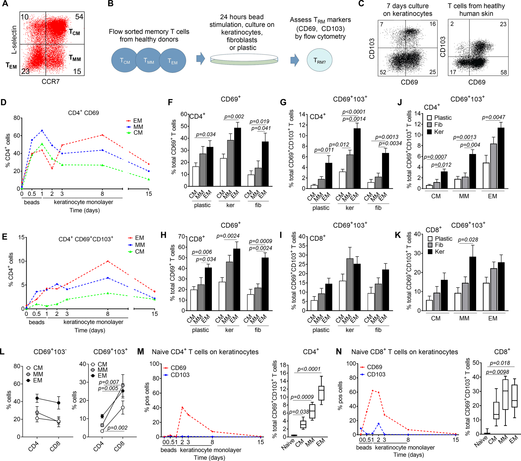
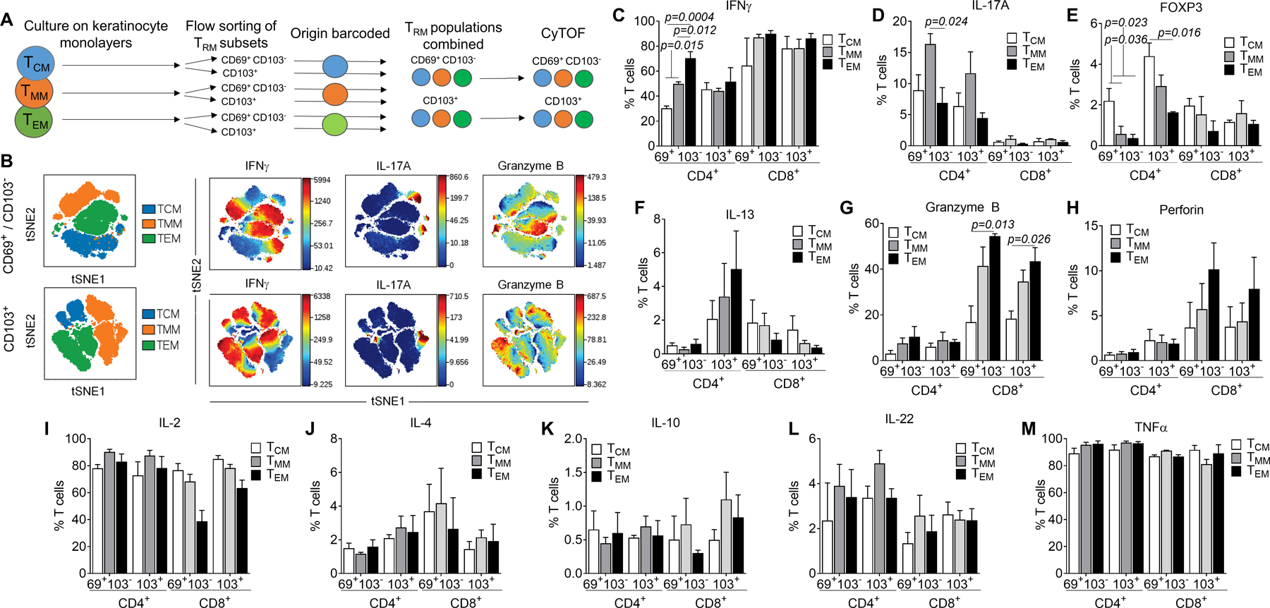
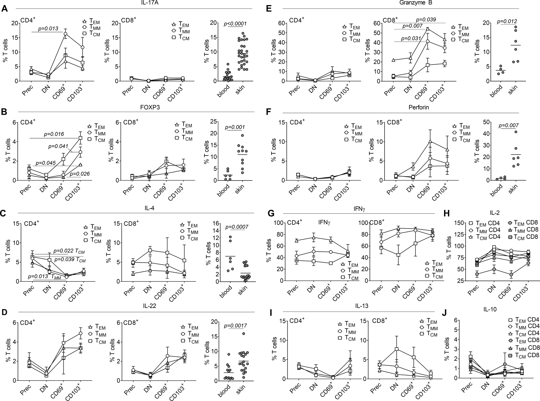
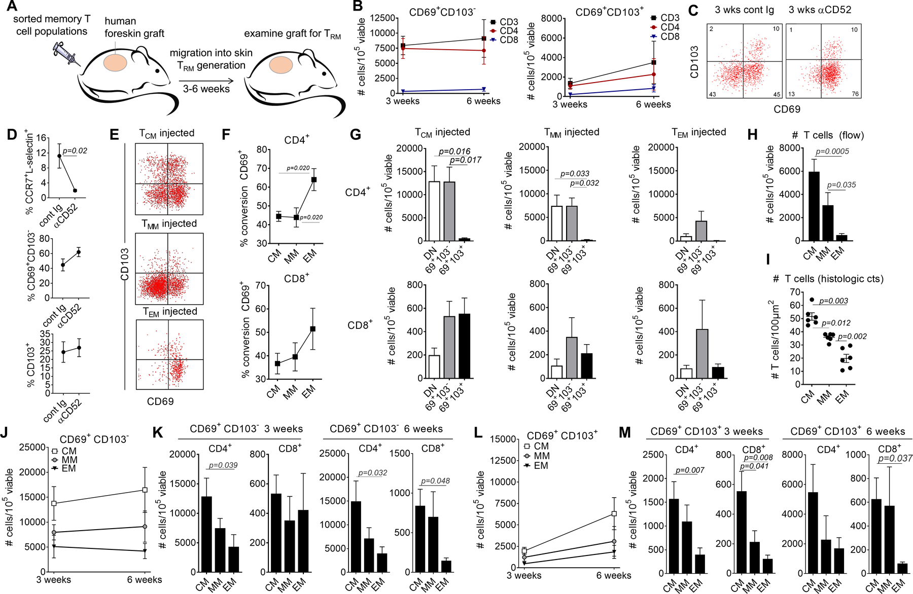
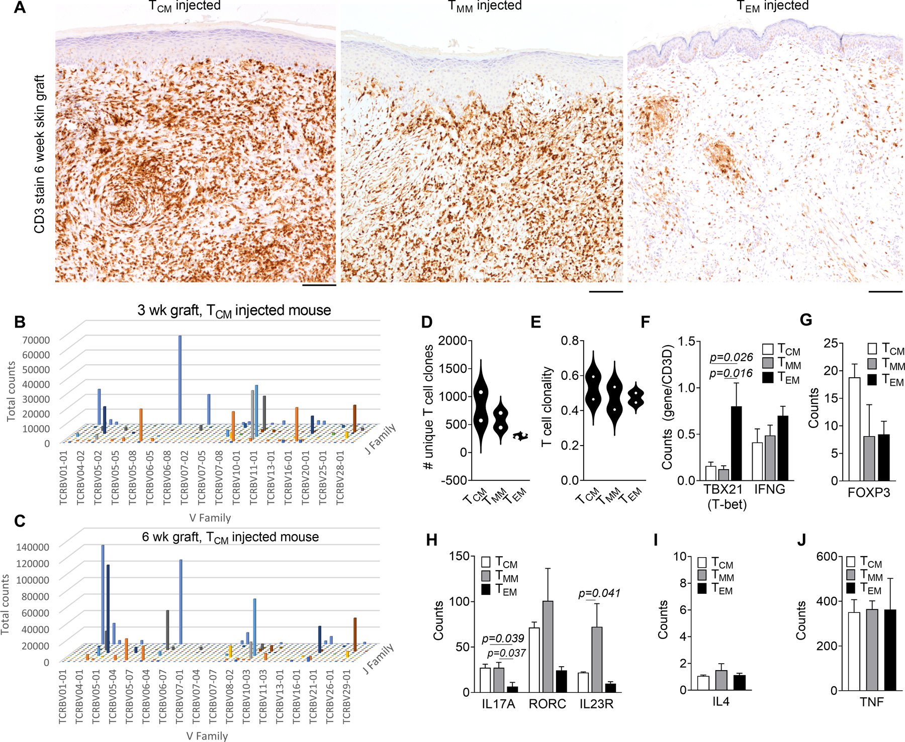
The circulating precursor cells that give rise to human resident memory T cells (TRM) are poorly characterized. We used an in vitro differentiation system and human skin-grafted mice to study TRM generation from circulating human memory T cell subsets. In vitro TRM differentiation was associated with functional changes, including enhanced IL-17A production and FOXP3 expression in CD4+ T cells and granzyme B production in CD8+ T cells, changes that mirrored the phenotype of T cells in healthy human skin. Effector memory T cells (TEM) had the highest conversion rate to TRM in vitro and in vivo, but central memory T cells (TCM) persisted longer in the circulation, entered the skin in larger numbers, and generated increased numbers of TRM. In summary, TCM are highly efficient precursors of human skin TRM, a feature that may underlie their known association with effective long-term immunity.


Conflict of interest statement

Competing interests. JTO is currently a full-time employee at Sanofi and holds company stock and/or stock options; there is no relevant conflict of interest with the work presented here. The other authors also report no competing interests.

Figures

Figure 1.
Figure 1.. Multiple human circulating memory T cell subsets can generate TRM in vitro.
(A) The gating strategy used for sorting of human peripheral blood CD45RA memory T cells into purified populations of CCR7L-selectin effector memory T cells (TEM), CCR7+L-selectin migratory memory T cells (TMM) and CCR7+L-selectin+ central memory T cells (TCM) is shown. (B) Experimental strategy for the study of TRM generation in vitro. Sorted purified T cell subsets from human blood were stimulated for 24 hours with an anti-CD3/CD2/CD28 beads, cultured on monolayers of keratinocytes, fibroblasts or on tissue culture plastic and analyzed by flow cytometry. (C) Seven days of culture on keratinocyte monolayers generated single positive CD69+CD103 and double positive CD69+CD103+ T cells (left panel), similar in phenotype to those observed in healthy human skin (right panel). (D, E) Time course of CD69 and CD103 acquisition in vitro after culture on keratinocyte monolayers. A representative culture is shown; similar results were obtained in a total of four donors. (F-I) All memory subsets tested gave rise to CD69+ T cells, some of which also upregulated CD103. Results for CD4 (top) and CD8 (bottom) T cells are shown after culture on tissue culture plastic (plastic), keratinocyte monolayers (ker) or fibroblast monolayers (fib). The mean and SEM of at least six donors are shown. (J, K) Culture on keratinocyte monolayers generated increased numbers of CD4 (J) and CD8 (K) T cells expressing TRM markers. The mean and SEM of a minimum of four donors are shown. (L) CD4+ and CD8+ T cells were equally efficient at up regulating CD69 (left panel) but CD8+ T cells were more efficient at generating double positive CD69+CD103+ T cells. The mean and SEM of at least four donors cultured on keratinocyte monolayers are shown. (M,N) Naïve T cells did not upregulate TRM markers in vitro. CD4+ (M) and CD8+ (N) CD45ROCD45RA+ naïve T cells were bead stimulated and cultured on keratinocyte monolayers then analyzed by flow cytometry. Similar results were obtained in a total of three naïve donors. Naïve T cells produced significantly fewer CD69+ CD103+ T cells (right panels); the mean and SEM of three naïve donors and six memory donors are shown. Significance was determined by a one-way ANOVA and Tukey’s post-hoc test for multiple comparisons (F-K, M,N) or by two-tailed T-tests (L).
Figure 2.
Figure 2.. TRM generated from distinct circulating T cell subsets differ in their functional characteristics.
(A) Mass cytometry was used to compare the functional characteristics of TRM generated in vitro from circulating precursor T cell populations. T cells expressing TRM markers were generated in vitro from each memory subset; CD69+ CD103 and CD103+ cells from each subset were pooled and analyzed together by mass cytometry. (B) Clustering suggested functional differences existed between TRM generated from distinct precursors. (C-M) The expression of each cytokine in the CD69+ CD103−− and CD103+ T cell populations for both CD4+ and CD8+ T cells are shown. The mean and SEM of experiments from three separate donors are shown. Individual tSNE plots and a list of antibodies used are included in Fig. S3 and Table SI. Significance was determined by a one-way ANOVA and Tukey’s post-hoc test for multiple comparisons.
Figure 3.
Figure 3.. Acquisition of TRM markers is accompanied by changes in T cell function.
Mass cytometry was utilized to compare production of cytokines and expression of FOXP3 in freshly isolated precursor T cell populations (Prec), vs. double negative (DN) CD69 CD103 T cells, single positive CD69+ CD103 (CD69+) and CD103+ (CD103+) T cells isolated after 1 day bead stimulation and 7 days of culture on keratinocyte monolayers. (A-J) Expression of the indicated cytokines is shown in each population, with comparisons to expression in blood CLA+ skin tropic T cells and T cells from healthy skin (A-F). The mean and SEM of mass cytometry results from three separate donors are shown. Significance was determined by a one-way ANOVA and Tukey’s post-hoc test for multiple comparisons (mass cytometry) or by two-tailed T-tests (flow cytometry studies of blood vs. skin).
Figure 4.
Figure 4.. Central memory T cells are the most efficient TRM precursors in vivo.
(A) An in vivo model using NSG mice grafted with human foreskin and infused with allogeneic human PBMC was utilized to study TRM formation in vivo in human skin. (B) TRM generated in this model were persistent. T cells were isolated from skin grafts three and six weeks after allogeneic PBMC infusion. The numbers of single positive CD69+ CD103 TRM (left panel) and double positive CD69+ CD103+ TRM (right panel) are shown for total CD3+, CD4+ and CD8+ T cells. The mean and SEM of 8 (3 weeks) and at least 4 (6 weeks) experiments are shown. (C,D) TRM generated in this model do not recirculate. Skin-grafted mice were treated i.p. with alemtuzumab or control Ig. (C) Example histograms from control Ig treated (cont Ig, left panel) and alemtuzumab treated mice (αCD52, right panel) are shown. Similar results were obtained in a total of four experiments, using four different blood donors. (D) The number of recirculating (CCR7+/L-selectin+) and TRM populations in mice treated with control Ig (cont Ig) and alemtuzumab (αCD52) are shown. The mean and SEM of four experiments using four different blood donors are shown. (E-G) All three memory subsets generated TRM in vivo. Foreskin-grafted mice were injected i.v. (1 mouse per precursor population) with flow purified populations of TCM, TMM and TEM and T cells were harvested from the skin graft and studied by flow cytometry after three weeks. A representative histogram is shown (E); similar results were obtained in a total of eight experiments, using eight different blood donor and a total of 24 mice. TEM had the highest percentage of conversion to CD69+ TRM (F,G). Cells were quantified as the number of T cells per 105 total viable cells (of all types) isolated from collagenase digested skin grafts. The mean and SEM of eight experiments are shown, using eight blood donors and 24 mice. (H, I) The total number of T cells in the skin grafts of injected mice are shown, as measured by quantitative flow cytometry (H) and histologic counts of T cells in CD3+ immunostained sections (I). The mean and SEM of 8 (H) and 6 (I) experiments are shown. (J-M) TCM generated the most TRM in vivo. The numbers of CD69+ CD103 (J,K) and CD69+ CD103+ TRM (L,M) among CD4+ (J,K) and CD8+(L,M) T cells are shown. The mean and SEM of experiments from eight donors (3 weeks) and at least four donors (6 weeks) are shown. Significance was determined by a one-way ANOVA and Tukey’s post-hoc test for multiple comparisons.
Figure 5.
Figure 5.. Diversity and functional characteristics of in vivo generated TRM.
(A) T cell infiltration into skin as assessed by CD3 immunostaining of grafts harvested six weeks after T cell injection is shown. Representative images from a total of eight experiments using eight different blood donors are shown. Scale bars = 100μm. (B,C) TCM-injected mice generated a diverse population of persistent TRM in skin. High-throughput TCR sequencing (HTS) results from 3 week (B) and 6 week (C) post infusion grafts from a single blood donor are shown. T cell clones identified in three weeks skin grafts were also identifiable at six weeks. (D, E) The number of unique T cell clones (D) and T cell clonality (E) in skin 6 weeks after infusion in TCM-injected mice is shown. The mean of experiments from two different blood donors are shown. (F-J) NanoString-based gene expression profiling was utilized to study functional characteristics of TRM generated from each memory subset. Results for type I (F), regulatory T cell (G), type 17 (H), type 2 (I) and TNFα (J) associated genes are shown. The mean and SEM from three experiments using three different blood donors, are shown. Significance was determined by a one-way ANOVA and Tukey’s post-hoc test for multiple comparisons.
Figure 6.
Figure 6.. TCM persist longer in the circulation, are skin tropic and replenish other memory T cell subsets.
(A-G) Circulating memory T cell precursor populations were analyzed by mass cytometry for the expression of cutaneous lymphocyte antigen (A), chemokine receptors (B), HLA-DR (C), BCL-2 (D), exhaustion markers PD-1 and Tim-3 (E,F) and Ki-67 (G). The mean and SEM of results from six different blood donors are shown. (H,I) TCM persist longer in the circulation. T cells from the blood and spleen of injected mice three weeks after infusion were analyzed by flow cytometry. The mean and SEM of six experiments using six different blood donors are shown. (I) TCM replenished other T cell subsets. The phenotype of T cells in blood three weeks after T cell infusion is shown. The mean and SEM of experiments using three different blood donors are shown. Significance was determined by a one-way ANOVA and Tukey’s post-hoc test for multiple comparisons.

References

    1. Park CO, Kupper TS, The emerging role of resident memory T cells in protective immunity and inflammatory disease. Nature medicine 21, 688–697 (2015). - PMC - PubMed
    1. Clark RA, Resident memory T cells in human health and disease. Sci Transl Med 7, 269rv261 (2015). - PMC - PubMed
    1. Mueller SN, Mackay LK, Tissue-resident memory T cells: local specialists in immune defence. Nature reviews Immunology 16, 79–89 (2016). - PubMed
    1. Sallusto F, Geginat J, Lanzavecchia A, Central Memory and Effector Memory T Cell Subsets: Function, Generation, and Maintenance. Annual review of immunology 22, 745–763 (2004). - PubMed
    1. Clark RA et al., Skin effector memory T cells do not recirculate and provide immune protection in alemtuzumab-treated CTCL patients. Science Translational Medicine 4, 117ra117 (2012). - PMC - PubMed

Publication types