. 2022 May 3;119(18):e2110085119.
doi: 10.1073/pnas.2110085119. Epub 2022 Apr 22.

The mechanism for ligand activation of the GPCR-G protein complex

Affiliations

The mechanism for ligand activation of the GPCR-G protein complex

Amirhossein Mafi et al. Proc Natl Acad Sci U S A. .

Abstract

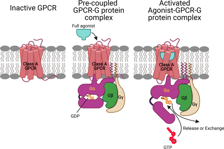
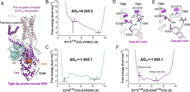
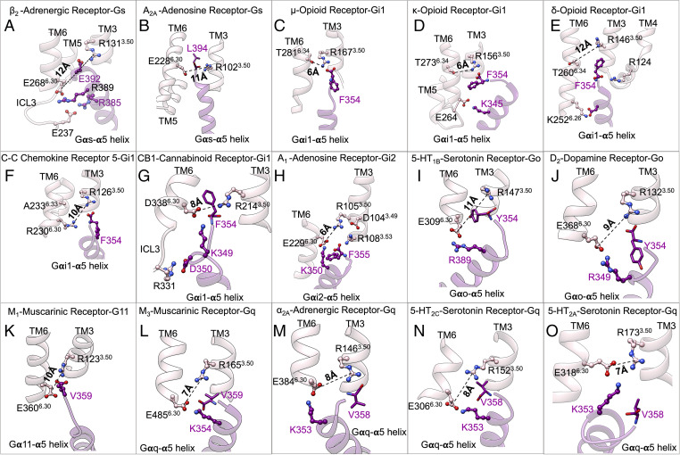
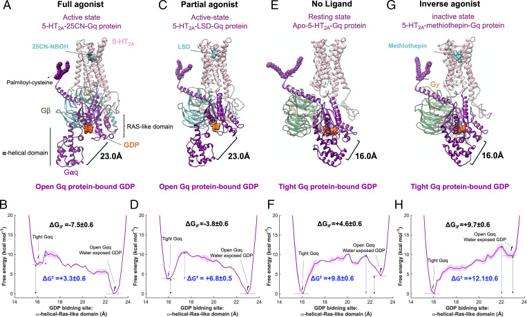
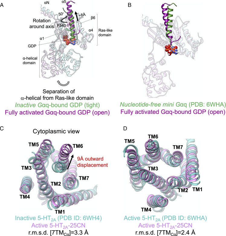
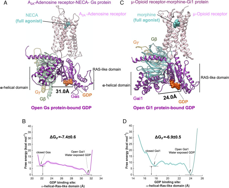
G protein–coupled receptors (GPCRs) activate cellular responses ranging from odorants to neurotransmitters. Binding an agonist leads to activation of a heterotrimeric G protein (GP) that stimulates external signaling. Unfortunately, the mechanism remains unknown. We show for 15 class A GPCRs, including opioids, adrenergics, adenosines, chemokines, muscarinics, cannabinoids, serotonins, and dopamines, that interaction of an inactive GP, including Gs, Gi, Go, G11, and Gq, to the inactive GPCR, containing the intracellular ionic lock between transmembrane (TM) helices 3 and 6, evolves exothermically to form a precoupled GPCR-GP complex with an opened TM3-TM6 and the GP-α5 helix partially inserted into the GPCR but not activated. We show that binding of agonist to this precoupled GPCR-GP complex causes the Gα protein to open into its active form, with the guanosine diphosphate exposed for signaling. This GP-first paradigm provides a strategy for developing selective agonists for GPCRs since it is the pharmacophore for the precoupled GPCR-GP complex that should be used to design drugs.

Keywords: G protein activation; adrenergic; biased agonists; molecular metadynamics; opioids.


Conflict of interest statement

The authors declare no competing interest.

Figures

Fig. 1.
Fig. 1.
GP-first mechanism of GP activation. Prior to ligand binding, the inactive GP interacts with the inactive GPCR to open the intracellular region by breaking the TM3-TM6 tight link to form a stable precoupled complex. This precoupled complex remains at this resting state until an agonist binds to the GPCR-GP complex to open the intracellular region of GPCR and the tightly coupled Gα-GDP complex to form the fully activated agonist-GPCR-GP complex with the GDP available for exchange or release.
Fig. 2.
Fig. 2.
The precoupled 5-HT2A-Gq protein complex. (A) Our energetically optimized precoupled complex of 5-HT2A-Gq protein-GDP. MetaD free energy of (B) R1733.50(CZ)-V358(C) and (C) E3186.30(CD)-K353(NZ). (D) Extensive engagement between the Gαq-α5 helix to the cytosolic end of TM3, and TM6 of 5-HT2A, along the Gq precoupling that breaks open the ionic lock between TM3 and TM6. (E) Detailed atomic interactions between 5-HT2A and Gαq-α5 helix in the precoupled state. (F) MetaD free energy of R1733.50(CZ)-E3186.30(CD) upon the formation of precoupling complex between 5-HT2A and Gq protein, which was estimated by performing an independent ∼600 ns metaD simulation. The metaD free energies were reweighted (51) for estimation of the free energy errors using the block averaging method.
Fig. 3.
Fig. 3.
Precoupled complexes of class A GPCR-GPs. Detailed atomic interactions between Gα5 helix and class A GPCRs in the precoupled state resulted from extensive metaD simulations for (A) β2-adrenergic receptor–Gs, (B) A2a adenosine–Gs, (C) μ-opioid–Gi1, (D) κ-opioid–Gi1, (E) δ-opioid–Gi1, (F) CCR5-chemokine–Gi1, (G) CB1-cannabinoid–Gi1, (H) A1-adenosine–Gi2 protein, (I) 5-HT1b-serotonin–Go, (J) D2-dopamine–Go, (K) M1-muscarinic–G11, (L) mouse M3-muscarinic–mouse Gq, (M) α2A-adrenergic–Gq, (N) 5-HT2c-serotonin–Gq, and (O) 5-HT2A-serotonin–Gq. The details of the calculations are represented in SI Appendix, Fig. S4 and Table S3. (CE) Adapted from figure 6A of ref. .
Fig. 4.
Fig. 4.
Ligand activation of precoupled 5-HT2A-Gq protein complex. Gq protein activation mediated by (A) 25CN-NBOH, a full agonist, and (C) LSD, a partial agonist, upon binding to the precoupled 5-HT2A-Gq protein complex. Gq protein inhibited activation caused by (E) absence of an agonist (apo-5-HT2A) and (G) methiothepin, an inverse agonist upon binding to the precoupled 5-HT2A-Gq protein complex. (B, D, F, and H) MetaD free energy of Gαq-bound GDP opening from its GDP binding site. Here, the distance between the AH domain (the center of mass of Cα for the residues 154 to 161 and 175 to 182) and the Ras-like domain (the center of mass of Cα for the residues 51 to 62), was considered for the free energy calculations. The weighted averages and the SDs were calculated for ΔGif for the converged period. The metaD free energies were reweighted (51) for estimation of the error, particularly energy barrier ΔG presented in B, D, F, and H, using the block averaging method.
Fig. 5.
Fig. 5.
Fully active state of 5-HT2A-Gq protein. Comparison of Gαq in our optimized fully active 25CN-NBOH-5-HT2A-Gq protein with (A) inactive Gαq protein-bound GDP and (B) fully active nucleotide free mini-Gαq subunit (54) resolved by cryo-EM. Comparison of the cytoplasmic region of 5-HT2A in our optimized fully active 25CN-NBOH-5-HT2A-Gq protein with (C) the inactive conformation (54) (Protein Data Bank [PDB]: 6WH4) resolved by X-ray crystallography and (D) the active conformation (54) (PDB: 6WHA) resolved by cryo-EM.
Fig. 6.
Fig. 6.
Ligand activation of precoupled GPCR-GP complex. Binding of a full agonist (A) NECA to the precoupled A2A adenosine receptor–Gs protein complex and (C) morphine to the precoupled μ-opioid receptor–Gi protein complex. MetaD free energy of (B) opening Gαs-bound GDP from its GDP binding site, distance between AH domain (center of mass of Cα for residues 69 to 204) and the Ras-like domain (center of mass of Cα for residues 49 to 65, 223 to 241, 294 to 303, and 369 to 374), and (D) opening Gαi-bound GDP from its GDP binding site (adapted from figure 6A from ref. 14), distance between AH domain (center of mass of Cα for residues 147 to 181) and Ras-like domain (center of mass of Cα for residues 42 to 59). The weighted averages and the SDs were calculated for ΔGif for the converged period. The errors associated with the free energy were calculated after reweighting (51) the free energies and using the block averaging method.

References

    1. Lefkowitz R. J., Seven transmembrane receptors: Something old, something new. Acta Physiol. (Oxf.) 190, 9–19 (2007). - PubMed
    1. Hauser A. S., Attwood M. M., Rask-Andersen M., Schiöth H. B., Gloriam D. E., Trends in GPCR drug discovery: New agents, targets and indications. Nat. Rev. Drug Discov. 16, 829–842 (2017). - PMC - PubMed
    1. Overington J. P., Al-Lazikani B., Hopkins A. L., How many drug targets are there? Nat. Rev. Drug Discov. 5, 993–996 (2006). - PubMed
    1. Fredriksson R., Lagerström M. C., Lundin L.-G., Schiöth H. B., The G-protein-coupled receptors in the human genome form five main families. Phylogenetic analysis, paralogon groups, and fingerprints. Mol. Pharmacol. 63, 1256–1272 (2003). - PubMed
    1. Clark A. J., The reaction between acetyl choline and muscle cells. J. Physiol. 61, 530–546 (1926). - PMC - PubMed

Publication types

LinkOut - more resources