. 2022 Apr 26;39(4):110729.
doi: 10.1016/j.celrep.2022.110729. Epub 2022 Apr 11.

Structural and functional impact by SARS-CoV-2 Omicron spike mutations

Affiliations

Structural and functional impact by SARS-CoV-2 Omicron spike mutations

Jun Zhang et al. Cell Rep. .

Abstract

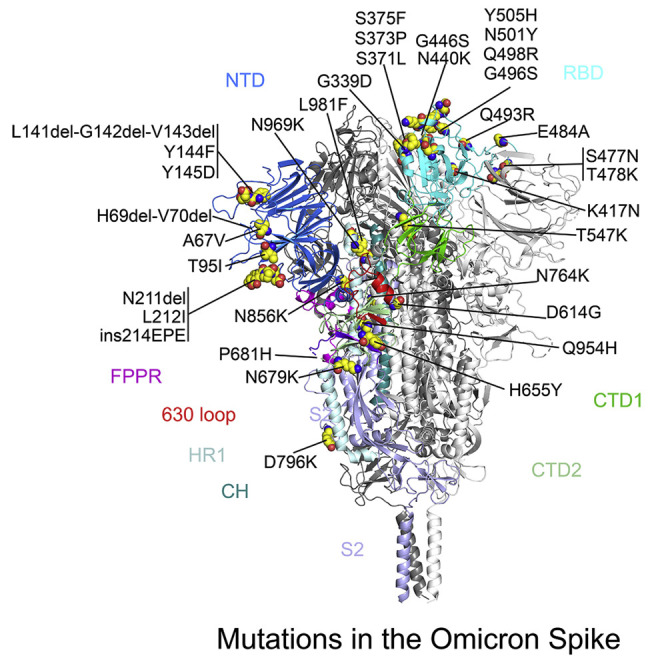
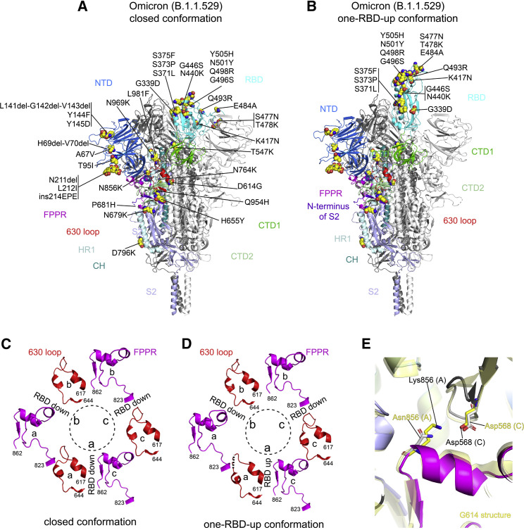
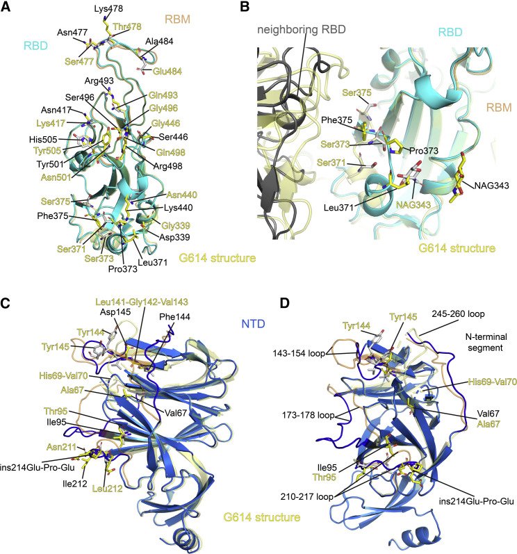
The Omicron variant of severe acute respiratory syndrome coronavirus 2 (SARS-CoV-2), bearing an unusually high number of mutations, has become a dominant strain in many countries within several weeks. We report here structural, functional, and antigenic properties of its full-length spike (S) protein with a native sequence in comparison with those of previously prevalent variants. Omicron S requires a substantially higher level of host receptor ACE2 for efficient membrane fusion than other variants, possibly explaining its unexpected cellular tropism. Mutations not only remodel the antigenic structure of the N-terminal domain of the S protein but also alter the surface of the receptor-binding domain in a way not seen in other variants, consistent with its remarkable resistance to neutralizing antibodies. These results suggest that Omicron S has acquired an extraordinary ability to evade host immunity by excessive mutations, which also compromise its fusogenic capability.

Keywords: CP: Molecular biology; SARS-CoV-2; cryo-EM; spike protein; structure.


Conflict of interest statement

Declaration of interests W.Y. serves on the scientific advisory boards of Hummingbird Bioscience and GO Therapeutics and is currently an employee of GV20 Therapeutics LLC. All other authors declare no competing interests.

Figures

None
Graphical abstract
Figure 1
Figure 1
Requirement of higher levels of ACE2 for efficient membrane fusion by the Omicron spike (A) Time course of cell-cell fusion mediated by various full-length S proteins, as indicated, with the target HEK293 cells transfected with 10 μg ACE2. (B) Time course of cell-cell fusion mediated by various full-length S proteins, as indicated, using HEK293 cells without exogenous ACE2. (C) Cell-cell fusion mediated by various full-length S proteins with HEK293 cells transfected with various levels (0–5 μg) of the ACE2 expression construct. (D) Cell-cell fusion mediated by various full-length S proteins expressed in HEK293 cells cotransfected with 5 μg furin expression construct and the ACE2-expressing target cells cotransfected with 5 μg TMPRSS2 expression construct. The experiments were performed in triplicates and repeated at least twice, with independent samples giving similar results. Error bars indicate the standard deviation calculated by the Excel STDEV function.
Figure 2
Figure 2
Antigenic properties of the purified full-length Omicron S protein Bio-layer interferometry (BLI) analysis of the association of prefusion S trimers derived from the G614 “parent” strain (B.1) and Omicron (B.1.1.529) variant with a soluble dimeric ACE2 construct and with a panel of antibodies representing five epitopic regions on the RBD and NTD (see Figure S5A and Tong et al., 2021). For ACE2 binding, purified ACE2 protein was immobilized to AR2G biosensors and dipped into the wells containing each purified S protein at various concentrations. For antibody binding, various antibodies were immobilized to anti-human immunoglobulin G (IgG) Fc capture (AHC) biosensors and dipped into the wells containing each purified S protein at different concentrations. Binding kinetics were evaluated using a 1:1 Langmuir model except for dimeric ACE2 and antibody G32B6 targeting the RBD-2, which were analyzed by a bivalent binding model. The sensorgrams are in black and the fits in red. Binding constants highlighted by underlines were estimated by steady-state analysis as described in the STAR Methods. Binding constants are also summarized here and in Table S1. N.D., not determined; RU, response unit. All experiments were repeated at least twice with essentially identical results.
Figure 3
Figure 3
cryo-EM structures of the full-length Omicron S protein (A) The structure of the closed prefusion conformation of the Omicron S trimer is shown in ribbon diagram with one protomer colored as NTD in blue, RBD in cyan, CTD1 in green, CTD2 in light green, S2 in light blue, the 630 loop in red, FPPR in magenta, HR1 in light blue, CH in teal, and the N-terminal segment of S2 in purple. All mutations in the Omicron variant, as compared with the original virus (Wuhan-Hu-1), are highlighted in sphere model. (B) The structure of the one-RBD-up conformation of the Omicron S trimer. (C) Structures, in the Omicron closed conformation, of segments (residues 617–644) containing the 630 loop (red) and segments (residues 823–862) containing the FPPR (magenta) from each of the three protomers (a–c). The position of each RBD is indicated. Dashed lines indicate gaps in the chain trace (disordered loops). ((D) Structures, in the Omicron one-RBD-up conformation, of segments (residues 617–644) containing the 630 loop (red) and segments (residues 823–862) containing the FPPR (magenta) from each of the three protomers (a–c). (E) Superposition of the structure of the Omicron S trimer in various colors with that of the G614 trimer in yellow aligned by S2, showing the region near the mutation N856K.
Figure 4
Figure 4
Structural impact of the mutations in the Omicron S (A) Superposition of the RBD structure of the Omicron S trimer in cyan with the RBD of the G614 S trimer in yellow. Locations of all 15 mutations in the RBD are indicated, and these residues are shown in stick model. The receptor-binding motif (RBM) is colored in orange in the G614 structure. (B) A close-up view of the RBD superposition in (A) to show the region near the mutations S371L, S373P, and S375F, including part of the neighboring RBD from another protomer. The mutated residues and the N-linked glycans at Asn343 are in stick model. NAG, N-acetylglucosamine. (C) Superposition of the NTD structure of the Omicron S trimer in blue with the NTD of the G614 S trimer in yellow. Locations of mutations A67V, T95I, Y144F, Y145D, and L212I; deletions H69del-V70del, L141del-G142del-V143del, and N211del; and an insertion ins214EPE are indicated, and these residues are shown in stick model. The N-terminal segment, 143–154, 173–187, 210–217, and 245–260 loops are rearranged between the two structures and highlighted in darker colors. (D) Another view of superposition of the NTD structure of the Omicron S trimer in blue with the NTD of the G614 S trimer in yellow.

References

    1. Adams P.D., Afonine P.V., Bunkoczi G., Chen V.B., Davis I.W., Echols N., Headd J.J., Hung L.W., Kapral G.J., Grosse-Kunstleve R.W., et al. PHENIX: a comprehensive Python-based system for macromolecular structure solution. Acta Crystallogr. D Biol. Crystallogr. 2010;66:213–221. - PMC - PubMed
    1. Adamson B., Sikka R., Wyllie A.L., Premsrirut P. Discordant SARS-CoV-2 PCR and rapid antigen test results when infectious: a December 2021 occupational case series. medRxiv. 2022 doi: 10.1101/2022.01.04.22268770. Preprint at. - DOI
    1. Altarawneh H., Chemaitelly H., Tang P., Hasan M.R., Qassim S., Ayoub H.H., AlMukdad S., Yassine H.M., Benslimane F.M., Khatib H.A.A., et al. Protection afforded by prior infection against SARS-CoV-2 reinfection with the Omicron variant. medRxiv. 2022 doi: 10.1101/2022.01.05.22268782. Preprint at. - DOI - PubMed
    1. Bosch B.J., van der Zee R., de Haan C.A., Rottier P.J. The coronavirus spike protein is a class I virus fusion protein: structural and functional characterization of the fusion core complex. J. Virol. 2003;77:8801–8811. - PMC - PubMed
    1. Cai Y., Zhang J., Xiao T., Lavine C.L., Rawson S., Peng H., Zhu H., Anand K., Tong P., Gautam A., et al. Structural basis for enhanced infectivity and immune evasion of SARS-CoV-2 variants. Science. 2021;373:642–648. - PMC - PubMed

Publication types

Substances

Supplementary concepts