Review
. 2022 Apr 21;82(8):1451-1466.
doi: 10.1016/j.molcel.2022.03.038.

Ribosome-associated quality-control mechanisms from bacteria to humans

Affiliations
Review

Ribosome-associated quality-control mechanisms from bacteria to humans

Sebastian Filbeck et al. Mol Cell. .

Abstract

Ribosome-associated quality-control (RQC) surveys incomplete nascent polypeptides produced by interrupted translation. Central players in RQC are the human ribosome- and tRNA-binding protein, NEMF, and its orthologs, yeast Rqc2 and bacterial RqcH, which sense large ribosomal subunits obstructed with nascent chains and then promote nascent-chain proteolysis. In canonical eukaryotic RQC, NEMF stabilizes the LTN1/Listerin E3 ligase binding to obstructed ribosomal subunits for nascent-chain ubiquitylation. Furthermore, NEMF orthologs across evolution modify nascent chains by mediating C-terminal, untemplated polypeptide elongation. In eukaryotes, this process exposes ribosome-buried nascent-chain lysines, the ubiquitin acceptor sites, to LTN1. Remarkably, in both bacteria and eukaryotes, C-terminal tails also have an extra-ribosomal function as degrons. Here, we discuss recent findings on RQC mechanisms and briefly review how ribosomal stalling is sensed upstream of RQC, including via ribosome collisions, from an evolutionary perspective. Because RQC defects impair cellular fitness and cause neurodegeneration, this knowledge provides a framework for pathway-related biology and disease studies.

Keywords: C-terminal tail; CAT tail; KLHDC10; LTN1; Listerin; NEMF; Pirh2; RQC; Rqc2; RqcH; alanine tail; quality control; ribosome; ribosome collision; ribosome stalling; translation.


Conflict of interest statement

Declaration of interests The authors declare no competing interests.

Figures
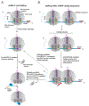
Figure 1:
Figure 1:. Ribosome stalling generates substrates for RQC.
Ribosome stalling at mRNA 3’ ends (A) and within open reading frames (ORFs) (B) are sensed differently, but both sensing systems lead to ribosomal splitting and generation of 60S subunits obstructed with peptidyl-tRNA, which are recognized by Rqc2/NEMF to initiate ribosome-associated quality control (RQC). Stalling within ORFs leads to ribosome collisions. The mechanisms in ‘A’ and ‘B’ may converge, with collided disomes causing trailing ribosomes to stall at mRNA 3’ ends, and conversely, with mRNA 3’ end-stlalled ribosomes leading to collisions if not resolved rapidly enough.
Figure 2:
Figure 2:. Bacterial proteolytic-targeting pathways elicited by ribosome stalling.
(A) Ribosomes stalled at mRNA 3’ ends are preferred substrates for trans-translation. (B) Ribosomes stalling within ORFs lead to collisions, which in bacteria are sensed by RqcU/MutS2 to elicit RQC. The RqcU ATPase promotes splitting of the stalled leading ribosome to generate 50S subunits obstructed with peptidyl-tRNA that serve as substrates for RQC. In both ‘A’ and ‘B’, the aberrant nascent-chains are tagged with C-terminal degrons (SsrA tag or Ala tail, respectively) and degraded by ClpXP or other proteases.
Figure 3:
Figure 3:. Aborted translation as a potential source of RQC substrates.
Environmental stresses, such as heat-shock or low Mg2+ concentration, can lead to ribosome destabilization during translation elongation and subsequent generation of large ribosomal subunits obstructed with peptidyl-tRNA, without the requirement for splitting factors.
Figure 4:
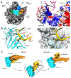
Figure 4:. Molecular architecture of eukaryotic and bacterial RQC complexes.
A, Structure of the human RQC complex as seen from the ribosomal subunit joining interface. The large ribosomal subunit (grey), a nascent-chain-linked P-site tRNA (pink), NEMF (blue) and Listerin (orange) are shown. B, Subunit architecture and domain scheme of RqcH orthologs. The structure and position of the NFACT-C domain specific to eukaryotes and archaea is not known to date. C, Structure of the bacterial RQC complex from B. subtilis. Coloring as in (A). Additionally, the bacteria-specific RQC component Hsp15/RqcP (green) and tRNAAla in the A-site (yellow) are shown. Figure based on EMD-2832 and EMD-11862.
Figure 5:
Figure 5:. tRNA recruitment and translocation during RqcH-mediated Ala tail synthesis in bacterial RQC.
A, Left: Structural overview of the bacterial RQC complex. Colored as in Fig. 1C. RqcH colored as in Fig. 1B. Right: Zoom on the nascent-chain-linked P-site tRNA (purple model), which is accommodated in a basic groove formed by the RqcH NFACT-N domain and Hsp15 (molecular surface representation; blue: positively charged, red: negatively charged). B, Structural basis for recruitment of tRNAAla by the NFACT-N domain. The tRNA anticodon nucleotides U34, G35 and C36 are accommodated in pockets of the NFACT-N domain. Coloring of atomic models as in (A). Zoom as indicated by the white box in (A). The NFACT-N domain is shown with (right) or without (left) the superposed molecular surface (transparent grey). C, Conformational changes of RqcH linked to tRNA hybrid state formation. The arrangement of the tRNA (yellow), the NFACT-N domain (blue) and the coiled-coil (orange) are shown for the decoding (left) and translocating state (right) of the Ala-tailing cycle. Middle: The two states are linked by a rotation of the NFACT-N domain and the associated tRNA. Figure based on EMD-11862, EMD-11864, PDB 7AQD and PDB 7AQC.
Figure 6:
Figure 6:. Model for the mammalian RQC pathway.
60S subunits obstructed with peptidyl-tRNA are recognized by NEMF, which elongates the nascent-chain C-termini with alanine residues (‘Ala tail’). Top: in the canonical RQC-L pathway, the E3 ligase Listerin/LTN1 is recruited for polyubiquitylation of the nascent-chain while the latter is still associated with the large ribosomal subunit. TCF25 directs the synthesis of ubiquitin Lys48-linked chains, which recruit the VCP complex (the binding mode of TCF25 has not been determined; we hypothesize that it spans from the intersubunit junction to the opening of the ribosomal exit tunnel, based on the ability of its yeast homolog, Rqc1, to selectively bind to 60S subunits in an Ltn1- and Rqc2-independent manner, as well as on the ability of TCF25 to influence ubiquitin chain synthesis). The nascent-chain is released by ANKZF1-mediated tRNA cleavage for VCP-mediated extraction and proteasomal degradation. Bottom: in the RQC-C pathway, presumably following peptidyl-tRNA hydrolysis by Ptrh1, the released nascent-chain is ubiquitylated by the E3 ligases Pirh2 or CRL2KLHDC10 for proteasomal degradation.

References

    1. Ahmed A, Wang M, Bergant G, Maroofian R, Zhao R, Alfadhel M, Nashabat M, AlRifai MT, Eyaid W, Alswaid A, et al. (2021). Biallelic loss-of-function variants in NEMF cause central nervous system impairment and axonal polyneuropathy. Hum. Genet 140, 579–592. - PubMed
    1. Bengtson MH, and Joazeiro CAP (2010). Role of a ribosome-associated E3 ubiquitin ligase in protein quality control. Nature 467, 470–473. - PMC - PubMed
    1. Brandman O, Stewart-Ornstein J, Wong D, Larson A, Williams CC, Li GW, Zhou S, King D, Shen PS, Weibezahn J, et al. (2012). A ribosome-bound quality control complex triggers degradation of nascent peptides and signals translation stress. Cell 151, 1042–1054. - PMC - PubMed
    1. Burby PE, and Simmons LA (2017). MutS2 Promotes Homologous Recombination in Bacillus subtilis. J. Bacteriol 199. - PMC - PubMed
    1. Burroughs AM, and Aravind L (2014). A highly conserved family of domains related to the DNA-glycosylase fold helps predict multiple novel pathways for RNA modifications. RNA Biol. 11, 360–372. - PMC - PubMed

Publication types

MeSH terms

LinkOut - more resources