Review
. 2022 Apr 21;82(8):1477-1491.
doi: 10.1016/j.molcel.2022.03.025.

Reshaping endoplasmic reticulum quality control through the unfolded protein response

Affiliations
Review

Reshaping endoplasmic reticulum quality control through the unfolded protein response

R Luke Wiseman et al. Mol Cell. .

Abstract

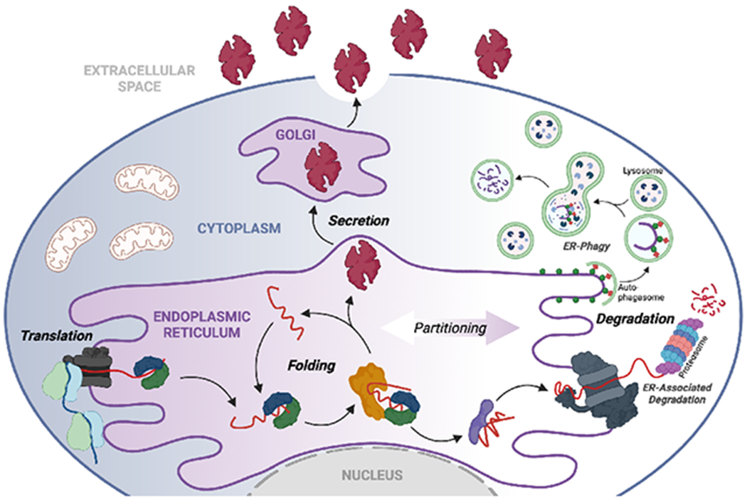
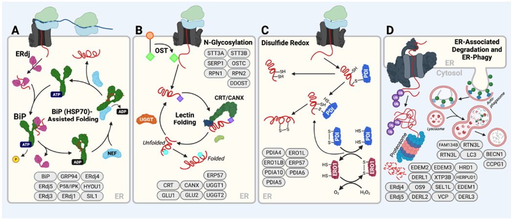
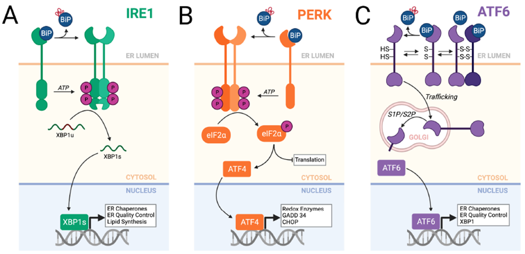
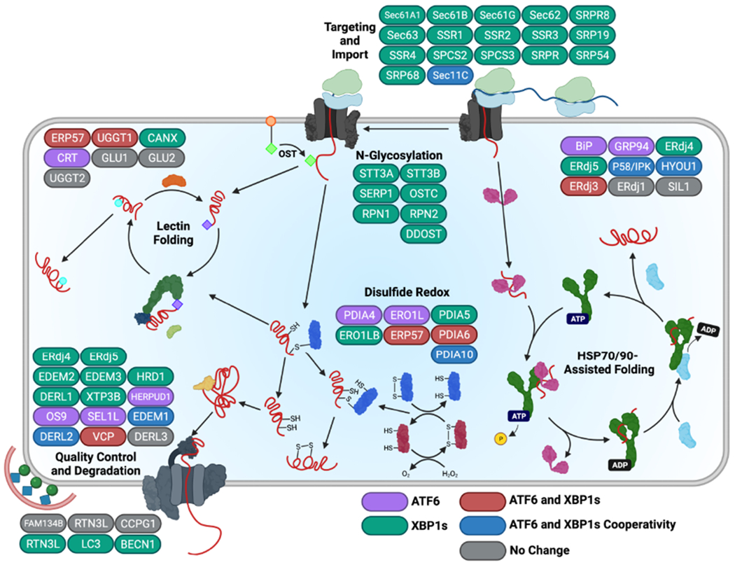
Endoplasmic reticulum quality control (ERQC) pathways comprising chaperones, folding enzymes, and degradation factors ensure the fidelity of ER protein folding and trafficking to downstream secretory environments. However, multiple factors, including tissue-specific secretory proteomes, environmental and genetic insults, and organismal aging, challenge ERQC. Thus, a key question is: how do cells adapt ERQC to match the diverse, ever-changing demands encountered during normal physiology and in disease? The answer lies in the unfolded protein response (UPR), a signaling mechanism activated by ER stress. In mammals, the UPR comprises three signaling pathways regulated downstream of the ER membrane proteins IRE1, ATF6, and PERK. Upon activation, these UPR pathways remodel ERQC to alleviate cellular stress and restore ER function. Here, we describe how UPR signaling pathways adapt ERQC, highlighting their importance for maintaining ER function across tissues and the potential for targeting the UPR to mitigate pathologies associated with protein misfolding diseases.

Keywords: ATF6; ER-associated degradation; ERAD; IRE1; PERK; XBP1s; amyloid; chaperone; loss-of-function disease; protein aggregation; protein misfolding disease.


Conflict of interest statement

Declaration of interests R.L.W. is an inventor on patents for IRE1/XBP1s and ATF6 activating compounds and is a scientific advisory board member and shareholder for Protego Biopharma, which has licensed UPR activating compounds for translational development. No other conflicts are identified.

Figures

Figure 1.
Figure 1.. ERQC is defined by the partitioning of proteins between ER protein folding/trafficking and degradation pathway.
Secretory proteins are co-translationally imported into the ER through the translocon channel where they immediately engage ER chaperones and folding factors. Through these interactions, proteins are assisted in attaining their folded conformation, allowing them to be trafficked to the Golgi and subsequently to downstream secretory environments such as the extracellular space. Proteins unable to attain a folded conformation within the ER are instead recognized by degradation factors and directed towards proteasomal or lysosomal degradation through ERAD or ER-phagy, respectively. Created with BioRender.com.
Figure 2.
Figure 2.. Core ERQC hubs that control the fate of nascent proteins entering the ER.
A. The BiP HSP70 chaperoning pathway. Substrates either bind directly to ATP-bound BiP or are delivered by ER-localized J-proteins (ERdjs), which stimulate BiP ATPase activity. This converts BiP to the ADP-bound conformation that has high affinity for substrates. Nucleotide exchange factors (NEFs) such as GRP170/HYOU1 then engage BiP to facilitate ADP-ATP exchange, returning BiP to the low-affinity ATP bound state and releasing the substrate for subsequent rounds of folding. Transfer of a client to ERdj family members like ERdj4 or ERdj5 can remove it from the folding cycle and transfer it for degradation. Components of the BiP chaperoning pathway are shown in ovals at the bottom of the panel. B. The Calnexin/Calreticulin lectin chaperoning pathway. Oligosaccharyl transferase (OST) appends a core glycan comprising Glc3-Man9-GlcNAc2 (green diamonds) to Asn at specific N-glycosyation site sequences (Asn-X-Ser/Thr). Two distal glucose residues are then removed from this core glycan by glucosidases, leaving a protein with a Glc-Man9-GlcNAc2 glycan (purple diamonds). Proteins possessing this singly glucosylated glycan are substrates for the lectin chaperones, calnexin (CANX) and calreticulin (CRT). Upon release from these chaperones, proteins can either fold into their native state or the final glucose of the N-glycan can be removed by glucosidases to leave a glycan comprising Man9-GlcNAc2 (blue circles) that cannot rebind CANX/CRT. This glucose-free glycan can then be either further trimmed by mannosidases that direct the protein to degradation or re-glucosylated by UGGT to allow further rounds of interactions with CANX or CRT. Select components of the Calnexin/Calreticulin lectin chaperoning pathway are shown at the bottom of the panel. C. Protein disulfide isomerase (PDI) activity in the ER. Oxidized PDIs form mixed disulfide bonds with Cys residues in substrate proteins that are then resolved by a second Cys in the substrate, creating a disulfide within the substrate protein. The resulting reduced PDI is then re-oxidized through the activity of ERO1. PDIs like ERdj5, a protein that also includes a J-domain, bind clients while in the reduced state causing the client disulfide to be transferred to ERdj5, thus serving to reduce the client for ERAD. Select components of the PDI folding pathway are shown. D. ER-associated degradation (ERAD) and ER-phagy. In ERAD, terminally misfolded proteins are directed to the ER retrotranslocon, which facilitates removal of non-native proteins from the ER to the cytosol. In the cytosol, these proteins are then ubiquitinated and subsequently degraded by the proteasome. Select components of the ERAD pathway are shown. In ER-phagy, misfolded proteins are directed to the lysosome for degradation by lysosomal hydrolases; select components are shown. Created with BioRender.com.
Figure 3.
Figure 3.. Mammalian UPR signaling pathways involved in adapting ERQC following an acute ER insult.
A. The IRE1/XBP1s signaling arm of the UPR. In response to ER stress, IRE1 is activated through a process involving dissociation of BiP from the IRE1 luminal domain. This leads to IRE1 oligomerization and subsequent activation of the IRE1 cytosolic kinase domain to promote autophosphorylation and allosteric activation of the IRE1 RNase. The activated IRE1 RNase adapts ERQC primarily through the non-canonical splicing of XBP1 mRNA, resulting in the production of the active transcription factor XBP1s. XBP1s activates expression of numerous genes involved in ERQC pathways including ER chaperones and degradation factors. B. The PERK arm of the UPR. In response to ER stress, BiP dissociates from the PERK luminal domain, allowing dimerization and autophosphorylation of the cytosolic kinase domain. This leads to selective PERK-dependent phosphorylation of eIF2α, which promotes both translational attenuation and selective activation of stress-responsive transcription factors such as ATF4. C. The ATF6 arm of the UPR. ATF6 is maintained in the ER as monomers and dimers, possessing intra- and inter-molecular disulfide bonds, that are bound to the ER chaperone BiP. ER stress promotes reduction of ATF6 disulfides and BiP dissociation, increasing populations of reduced ATF6 monomers that traffic to the Golgi where they are processed by site 1 (S1P) and site 2 (S2P) proteases. This releases the active ATF6 bZIP transcription factor domain, which localizes to the nucleus and induces expression of stress-responsive genes primarily involved in ERQC. Created with BioRender.com.
Figure 4.
Figure 4.. XBPIs and/or ATF6 activation promote distinct remodeling of ERQC pathways.
XBPIs and ATF6 induce expression of specific, but overlapping, sets of ERQC factors that differentially impact ER function. ERQC genes primarily up-regulated by XBP1s are shown in green, while those primarily induced by ATF6 are depicted in purple. Genes targeted by both XBP1s and ATF6 to similar extents are shown in red, whereas genes regulated cooperatively by ATF6 and XBP1s activation are in blue. Image was adapted from (Shoulders et al., 2013) where the individual expression of ERQC factors induced by XBP1s and/or ATF6 activation in the absence of ER stress was measured in HEK293 cells. Created with BioRender.com.

References

    1. Adachi Y, Yamamoto K, Okada T, Yoshida H, Harada A, and Mori K (2008). ATF6 is a transcription factor specializing in the regulation of quality control proteins in the endoplasmic reticulum. Cell Struct Funct 33, 75–89. - PubMed
    1. Adams BM, Canniff NP, Guay KP, Larsen ISB, and Hebert DN (2020). Quantitative glycoproteomics reveals cellular substrate selectivity of the ER protein quality control sensors UGGT1 and UGGT2. Elife 9. - PMC - PubMed
    1. Adams BM, Oster ME, and Hebert DN (2019). Protein Quality Control in the Endoplasmic Reticulum. Protein J 38, 317–329. - PMC - PubMed
    1. Adamson B, Norman TM, Jost M, Cho MY, Nunez JK, Chen Y, Villalta JE, Gilbert LA, Horlbeck MA, Hein MY, et al. (2016). A Multiplexed Single-Cell CRISPR Screening Platform Enables Systematic Dissection of the Unfolded Protein Response. Cell 167, 1867–1882. - PMC - PubMed
    1. Amin-Wetzel N, Saunders RA, Kamphuis MJ, Rato C, Preissler S, Harding HP, and Ron D (2017). A J-Protein Co-chaperone Recruits BiP to Monomerize IRE1 and Repress the Unfolded Protein Response. Cell 171, 1625–1637 e1613. - PMC - PubMed

Publication types

LinkOut - more resources