. 2022 Sep;77(3):619-631.
doi: 10.1016/j.jhep.2022.04.010. Epub 2022 Apr 19.

Lack of VMP1 impairs hepatic lipoprotein secretion and promotes non-alcoholic steatohepatitis

Affiliations

Lack of VMP1 impairs hepatic lipoprotein secretion and promotes non-alcoholic steatohepatitis

Xiaoxiao Jiang et al. J Hepatol. 2022 Sep.

Abstract

Background & aims: Vacuole membrane protein 1 (VMP1) is an endoplasmic reticulum (ER) transmembrane protein that regulates the formation of autophagosomes and lipid droplets. Recent evidence suggests that VMP1 plays a critical role in lipoprotein secretion in zebra fish and cultured cells. However, the pathophysiological roles and mechanisms by which VMP1 regulates lipoprotein secretion and lipid accumulation in non-alcoholic fatty liver disease (NAFLD) and non-alcoholic steatohepatitis (NASH) are unknown.

Methods: Liver-specific and hepatocyte-specific Vmp1 knockout mice as well as Vmp1 knock-in mice were generated by crossing Vmp1flox or Vmp1KI mice with albumin-Cre mice or by injecting AAV8-TBG-cre, respectively. Lipid and energy metabolism in these mice were characterized by metabolomic and transcriptome analyses. Mice with hepatic overexpression of VMP1 who were fed a NASH diet were also characterized.

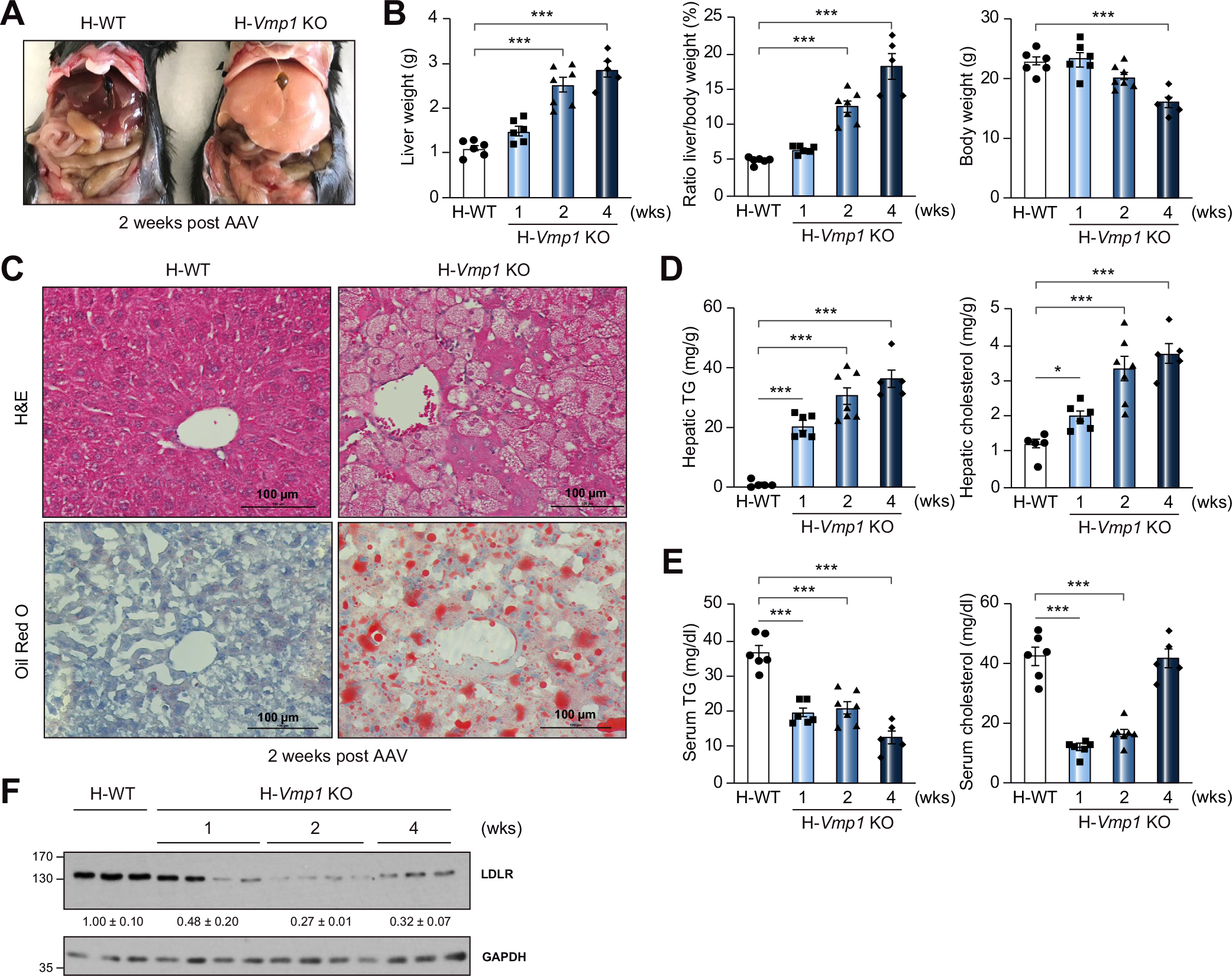
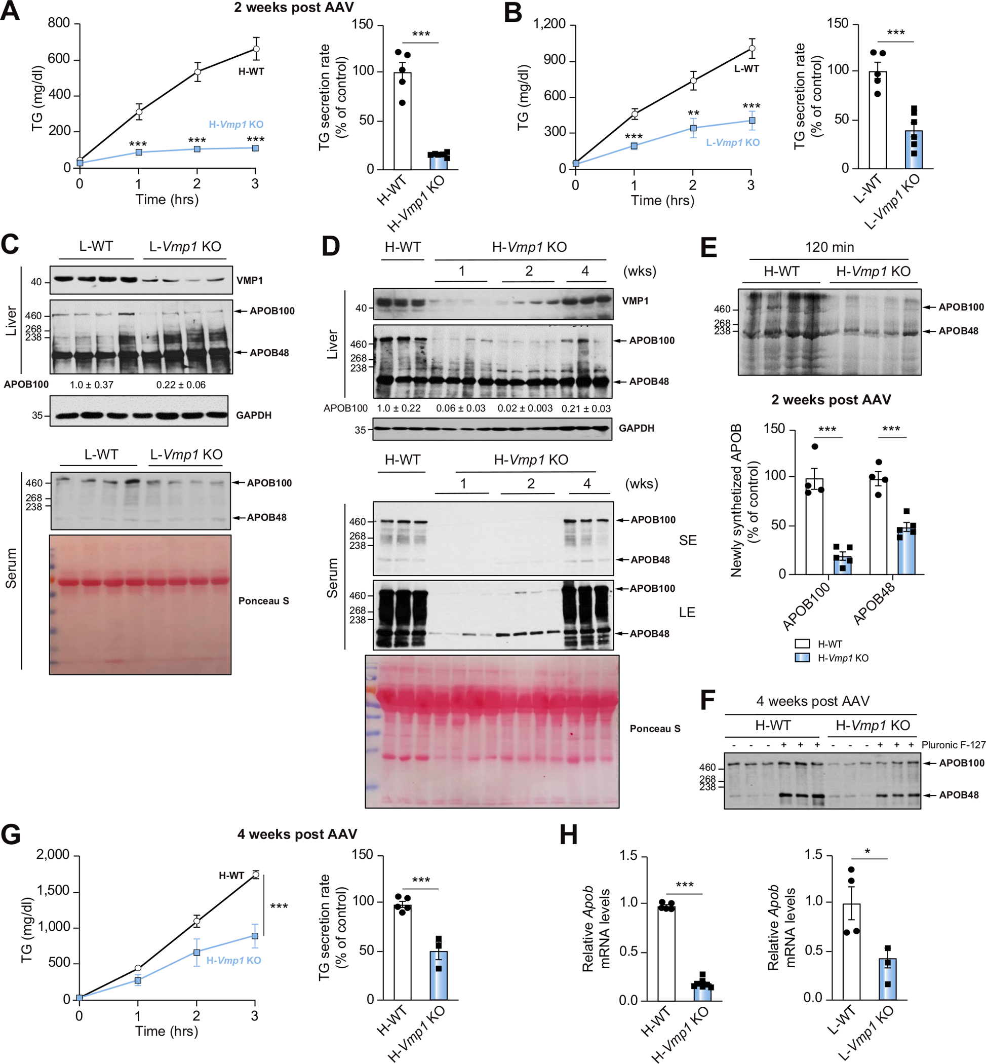
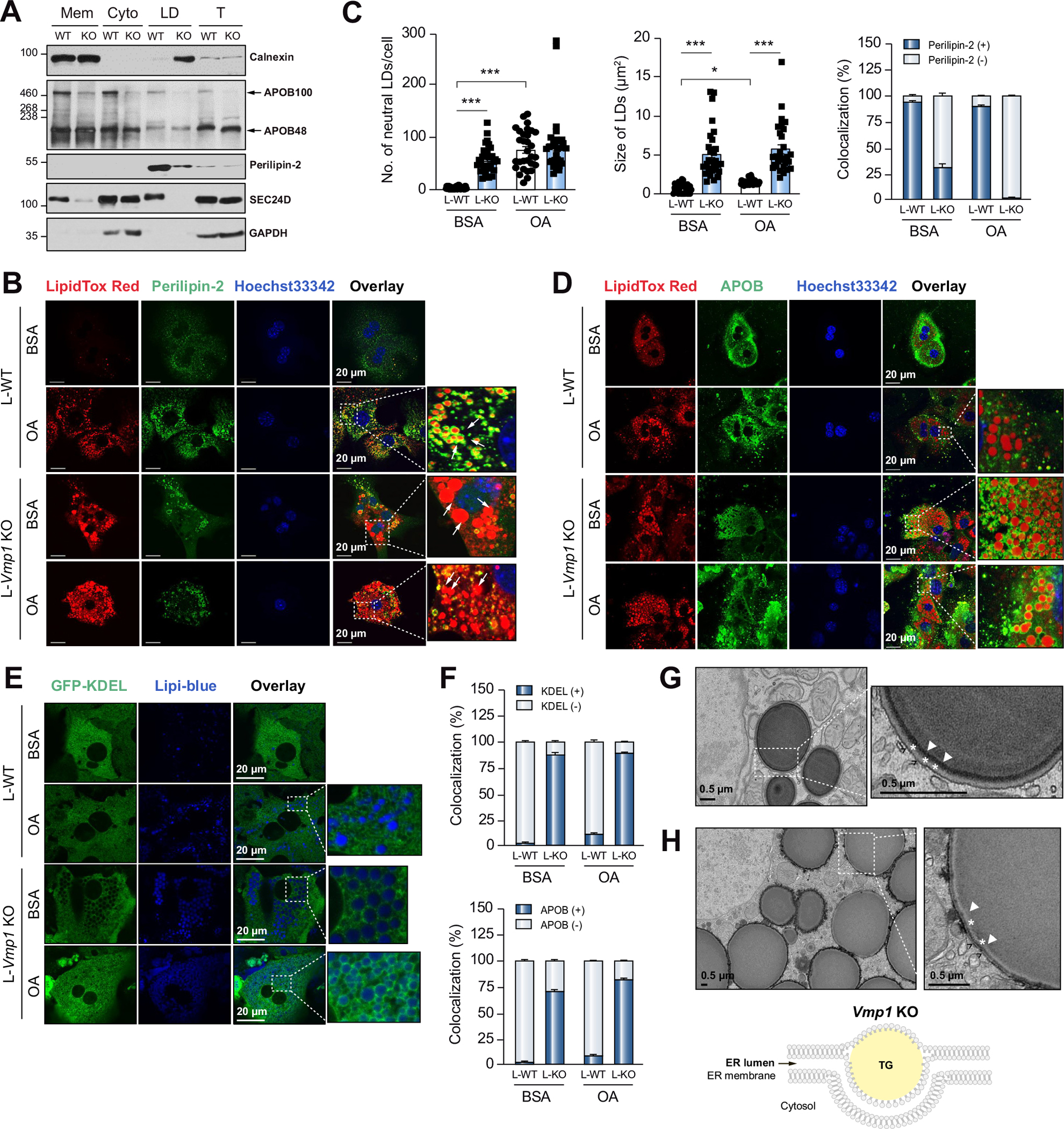
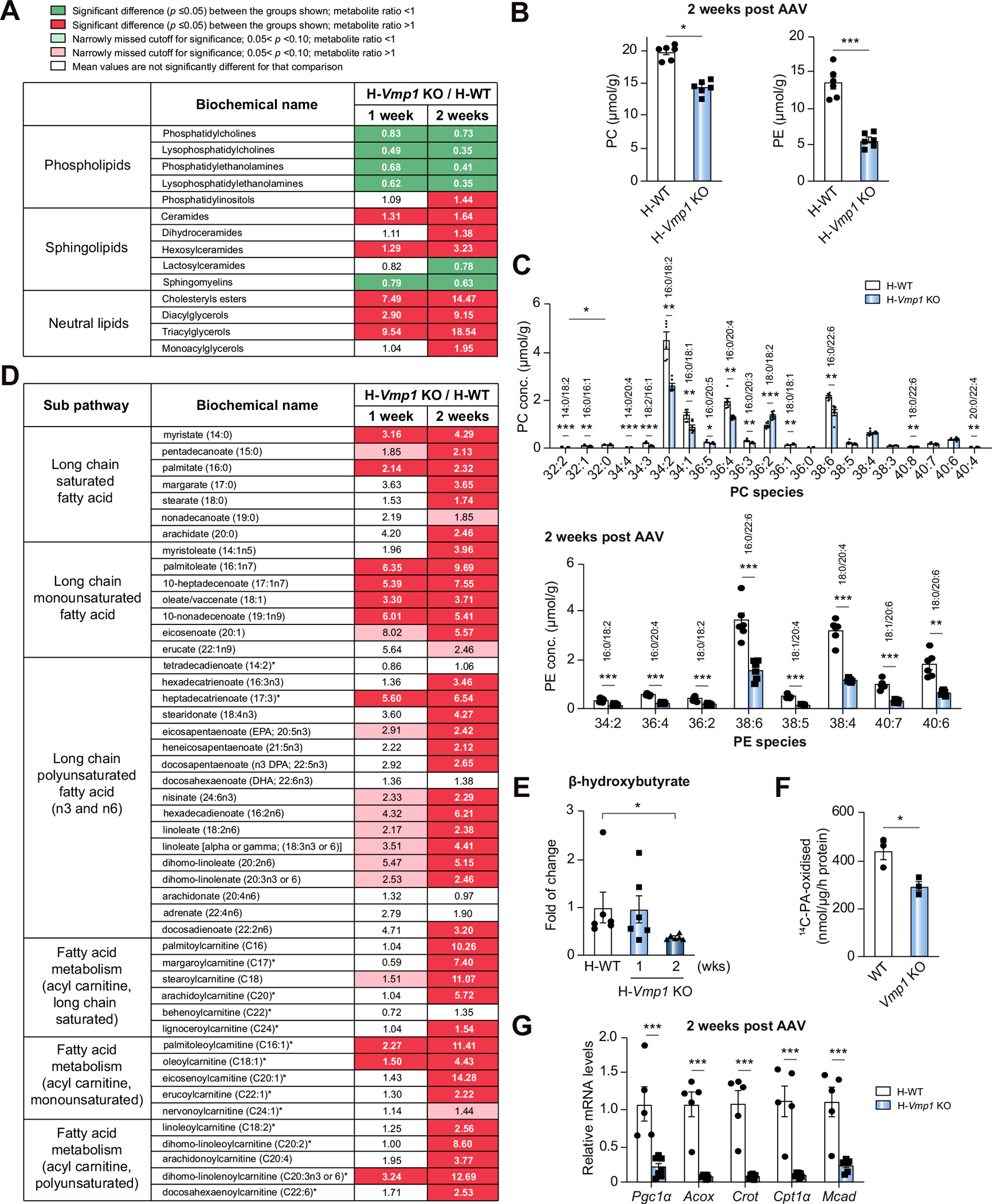
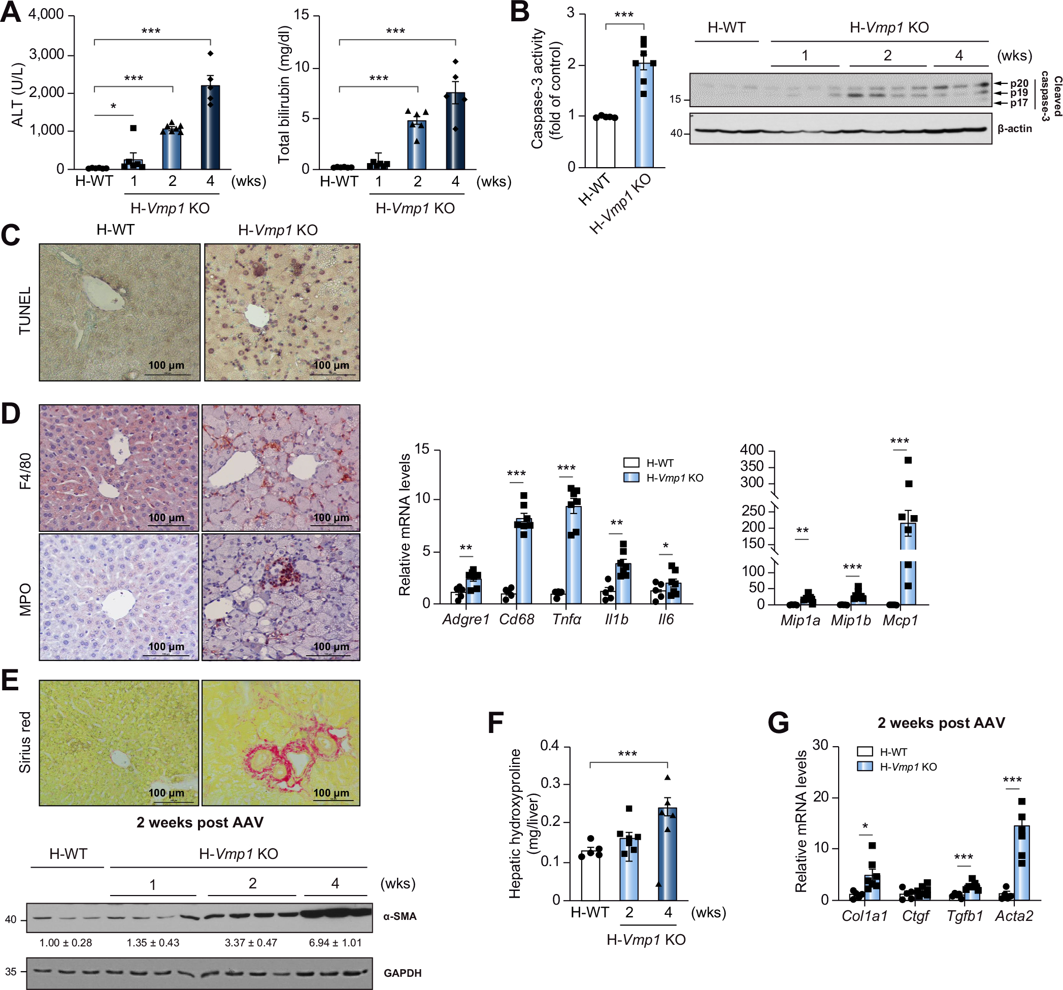
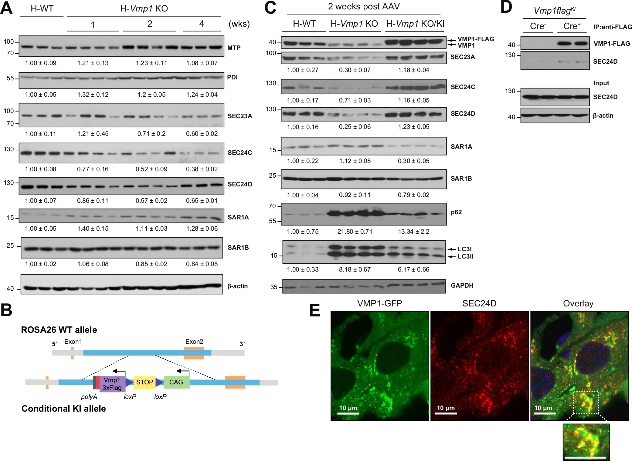
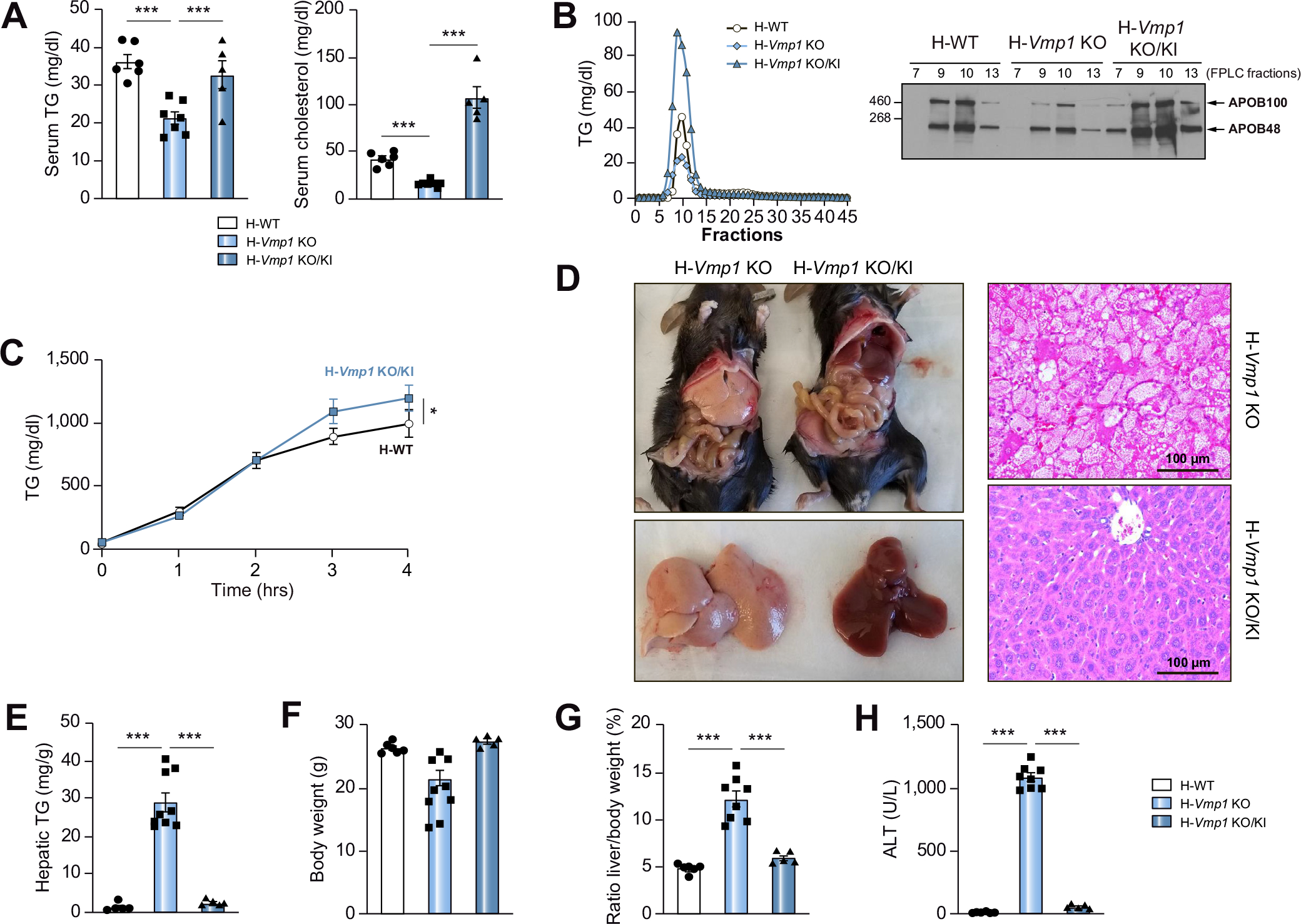
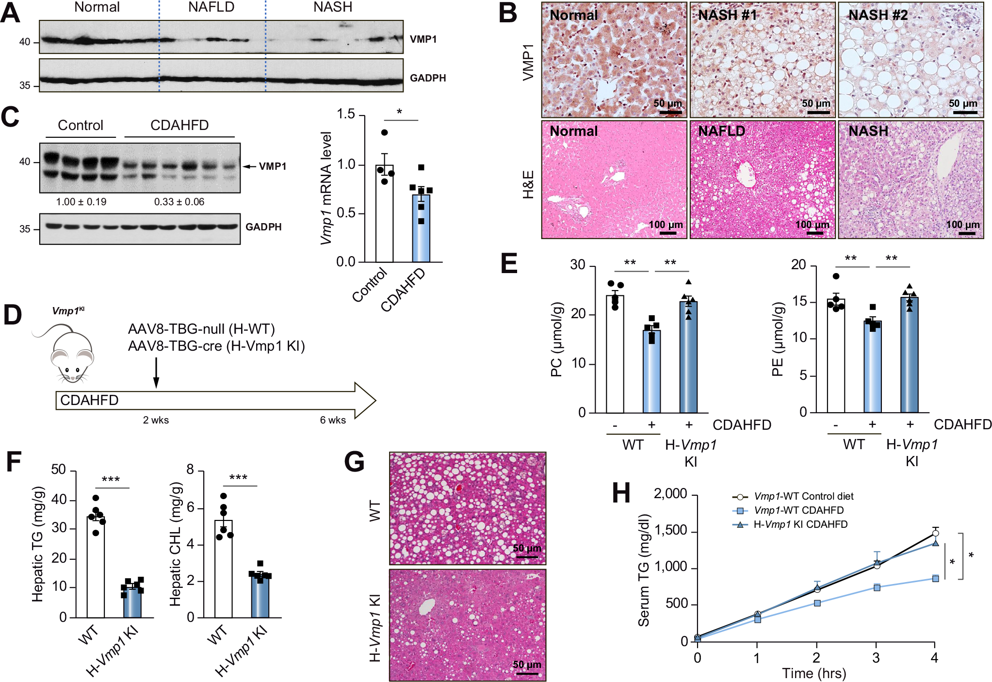
Results: Hepatocyte-specific deletion of Vmp1 severely impaired VLDL secretion resulting in massive hepatic steatosis, hepatocyte death, inflammation and fibrosis, which are hallmarks of NASH. Mechanistically, loss of Vmp1 led to decreased hepatic levels of phosphatidylcholine and phosphatidylethanolamine as well as to changes in phospholipid composition. Deletion of Vmp1 in mouse liver also led to the accumulation of neutral lipids in the ER bilayer and impaired mitochondrial beta-oxidation. Overexpression of VMP1 ameliorated steatosis in diet-induced NASH by improving VLDL secretion. Importantly, we also showed that decreased liver VMP1 is associated with NAFLD/NASH in humans.

Conclusions: Our results provide novel insights on the role of VMP1 in regulating hepatic phospholipid synthesis and lipoprotein secretion in the pathogenesis of NAFLD/NASH.

Lay summary: Non-alcoholic fatty liver disease and its more severe form, non-alcoholic steatohepatitis, are associated with a build-up of fat in the liver (steatosis). However, the exact mechanisms that underly steatosis in patients are not completely understood. Herein, the authors identified that the lack of a protein called VMP1 impairs the secretion and metabolism of fats in the liver and could therefore contribute to the development and progression of non-alcoholic fatty liver disease.

Keywords: NAFLD; VLDL; autophagy; endoplasmic reticulum; liver injury.


Conflict of interest statement

Conflict of interest The authors who have taken part in this study declare that they have nothing to disclose. Please refer to the accompanying ICMJE disclosure forms for further details.

Figures

Fig. 1.
Fig. 1.. Loss of hepatic VMP1 leads to steatosis in mice.
(A) Representative images of 8–10-week-old Vmp1flox mice at 2 weeks post AAV8-TBG-null (H-WT) or AAV8-TBG-cre (H-Vmp1 KO) injection. (B) Mouse liver, liver/body weight ratio and body weight. (C) H&E and Oil Red O staining of liver tissues from H-WT and H-Vmp1 KO mice. Hepatic (D) and serum (E) TG and cholesterol were quantified. (F) Total liver lysates were subjected to immunoblot analysis. Data represent mean ± SEM (n = 5–7). ***p <0.001 (One-way ANOVA with Holm-Sidak post hoc test). KO, knockout; TG, triglyceride; WT, wild-type.
Fig. 2.
Fig. 2.. Reduced TG and lipoprotein secretion in hepatocyte-specific and liver-specific Vmp1 KO mice.
(A) H-WT and H-Vmp1 KO mice or (B) one-month-old L-WT and L-Vmp1 KO mice were injected with tyloxapol and serum TG concentrations and secretion rates were measured. Total liver lysates and serum from L-WT and L-Vmp1 KO mice (C) or H-WT and H-Vmp1 KO mice (D) were subjected to immunoblot analysis. (E) Mice were fasted for 4–5 hours followed by injection via tail vein with 1 mg/g Pluronic F-127 and 15 mCi/kg of 35S-methionine labeling mix. Newly synthesized APOB was quantified 120 minutes later in H-WT and H-Vmp1 KO mouse sera. (F) Immunoblot analysis of serum APOB. (G) TG concentrations and secretion rates were measured in H-WT and H-Vmp1 KO mice at 4 weeks post AAV. (H) qPCR analysis of hepatic Apob mRNA. Data represent mean ± SEM (n = 4–7). *p <0.05; **p <0.01; ***p <0.001 (Unpaired Student’s t test). APO, apolipoprotein; LE, long exposure; SE, short exposure.
Fig. 3.
Fig. 3.. Loss of hepatic VMP1 causes accumulation of lipids in the endoplasmic reticulum bilayer.
(A) Liver subcellular fractions were subjected to immunoblot analysis. (B) Primary hepatocytes were treated with BSA or OA (200 μM) for 6 hours and stained with perilipin-2 and LipidTOX Red followed by confocal microscopy. White arrows indicate LDs. (C) Total number, size of LDs, and colocalization of neutral lipids with perilipin-2 were quantified (≥30 cells of 3 independent experiments). (D) Primary hepatocytes were stained with APOB and LipidTOX Red followed by confocal microscopy. (E) Primary hepatocytes were infected with Ad-ssRFP-GFP-KDEL and stained with Lipi-blue followed by confocal microscopy. (F) Colocalization of LDs with APOB and KDEL were quantitated (≥20 cells of 2 independent experiments). Representative EM images of primary hepatocyte from L-Vmp1 KO mice (G) or H-Vmp1 KO mouse livers at 2 weeks post AAV (H). *p <0.05; ***p <0.001 (One-way ANOVA with Holm-Sidak post hoc test). BSA, bovine serum albumin; Cyto, cytosol; ER, endoplasmic reticulum; LD, lipid droplet; Mem, membrane; OA, oleic acid; T, total lysate.
Fig. 4.
Fig. 4.. Loss of VMP1 in hepatocytes leads to reduced phospholipids and impaired fatty acid β-oxidation.
(A) Heatmaps of phospholipids, sphingolipids and neutral lipids, (B) total PC and PE, and (C) PC and PE species by metabolomics analysis of H-WT and H-Vmp1 KO mouse livers at 2 weeks. (D) Heatmaps of fatty acid metabolism and (E) β-hydroxybutyrate changes by metabolomics analysis. (n = 6). (F) FAO was measured in primary hepatocytes (n = 3 independent experiments). (G) qPCR analysis of hepatic FAO gene expression in mouse livers. Data represent mean ± SEM (n = 5–7). *p <0.05; **p <0.01; ***p <0.001 (Unpaired Student’s t test for 2 group comparison or one-way ANOVA with Holm-Sidak post hoc test for multigroup comparison). PC, phosphatidylcholine; PE, phosphatidylethanolamine.
Fig. 5.
Fig. 5.. H-Vmp1 KO mice develop NASH.
(A) Serum ALT and bilirubin of H-WT and H-Vmp1 KO mice were measured. (B) Caspase-3 activity and cleaved caspase-3 were analyzed using total liver lysates. (C) TUNEL staining and (D) immunohistochemistry staining for F4/80 and MPO in mouse livers as well as qPCR analysis of hepatic inflammatory genes. (E) Sirius red staining and immunoblot analysis of α-SMA in mouse livers. (F) Hepatic hydroxyproline and (G) qPCR analysis of fibrotic gene expression was quantified. Data represent mean ± SEM (n = 5–7). *p <0.05; **p <0.01; ***p <0.001 (Unpaired Student’s t test for 2 group comparison or one-way ANOVA with Holm-Sidak post hoc test for multigroup comparison). ALT, alanine aminotransferase.
Fig. 6.
Fig. 6.. Reduced COPII in hepatic Vmp1 KO mice.
(A) Total liver lysates of indicated mice were subjected to immunoblot analysis. (B) Strategy for generating conditional Vmp1KI mice. (C) Total liver lysates of indicated mice were subjected to immunoblot analysis. (D) Immunoprecipitation assay for VMP1 and SEC24D in mouse livers. (E) Vmp1flox mice were injected with AAV8-TBG-cre for 1 week followed by injecting Ad-Vmp1-Gfp for another 2 weeks. Immunostaining for SEC24D was performed and the colocalization of VMP1-GFP and SEC24D was assessed by confocal microscopy. KI, knock-in.
Fig. 7.
Fig. 7.. Restoration of VMP1 in Vmp1 KO mice improves VLDL secretion and ablates hepatic steatosis and liver injury.
(A) Serum TG and cholesterol were measured in H-WT, H-Vmp1 KO and H-Vmp1 KO/KI mice at 2 weeks post AAV. (B) Mice were fasted for 4 hours followed by Pluronic F-127 injection for another 4 hours. Pooled serum samples were subjected to FPLC analysis of lipoproteins (n = 5), and each corresponding fraction was subjected to immunoblot analysis for APOB. (C) VLDL secretion was assessed in mice after VMP1 restoration. (D) Representative images of gross livers, H&E and Oil Red O staining of mouse livers. (E-H) Hepatic TG (E), body weight (F), liver/body weight ratio (G) and ALT (H) were quantified. Data represent mean ± SEM (n = 5–7). *p <0.05; ***p <0.001 (Unpaired Student’s t test for 2 group comparison or one-way ANOVA with Holm-Sidak post hoc test for multigroup comparison).
Fig. 8.
Fig. 8.. Decreased VMP1 in human NAFLD and overexpression of VMP1 alleviates diet-induced steatosis in mice.
(A) Total lysates from human livers were subjected to immunoblot analysis. (B) Representative images of VMP1 IHC and H&E staining of normal and NAFLD/NASH patient livers. (C) H-WT mice were fed with CDAHFD for 6 weeks. Protein and mRNA levels of VMP1 in mouse livers were measured by immunoblot and qPCR analysis. (D) Scheme of CDAHFD-induced NASH in mice. (E) Hepatic concentrations of PC and PE, (F) TG and cholesterol and (G) H&E staining of liver tissues. (H) VLDL secretion was assessed in WT and VMP1 KI mice. Data represent mean ± SEM (n = 5–7). *p <0.05; ***p <0.001 (Unpaired Student’s t test for 2 group comparison or one-way ANOVA with Holm-Sidak post hoc test for multigroup comparison). NAFLD, non-alcoholic fatty liver disease; NASH, non-alcoholic steatohepatitis.

References

    1. Cohen JC, Horton JD, Hobbs HH. Human fatty liver disease: old questions and new insights. Science 2011;332:1519–1523. - PMC - PubMed
    1. Alves-Bezerra M, Cohen DE. Triglyceride metabolism in the liver. Compr Physiol 2017;8:1–8. - PMC - PubMed
    1. Davidson NO, Shelness GS. APOLIPOPROTEIN B: mRNA editing, lipoprotein assembly, and presecretory degradation. Annu Rev Nutr 2000;20:169–193. - PubMed
    1. Samuel VT, Shulman GI. Nonalcoholic fatty liver disease as a nexus of metabolic and hepatic diseases. Cell Metab 2018;27:22–41. - PMC - PubMed
    1. Dusetti NJ, Jiang Y, Vaccaro MI, Tomasini R, Azizi Samir A, Calvo EL, et al. Cloning and expression of the rat vacuole membrane protein 1 (VMP1), a new gene activated in pancreas with acute pancreatitis, which promotes vacuole formation. Biochem Biophys Res Commun 2002;290:641–649. - PubMed

Publication types