. 2022 May:94:107020.
doi: 10.1016/j.ijscr.2022.107020. Epub 2022 Apr 5.

Renal cell carcinoma with early skin metastasis and partial response on tyrosine kinase inhibitor: A case report

Affiliations

Renal cell carcinoma with early skin metastasis and partial response on tyrosine kinase inhibitor: A case report

Agus Rizal Ardy Hariandy Hamid et al. Int J Surg Case Rep. 2022 May.

Abstract

Introduction and importance: Renal cell carcinoma (RCC) skin metastasis is a rare disease. However, there are no data on the effect of a Tyrosine Kinase Inhibitor (TKI) on its treatment.

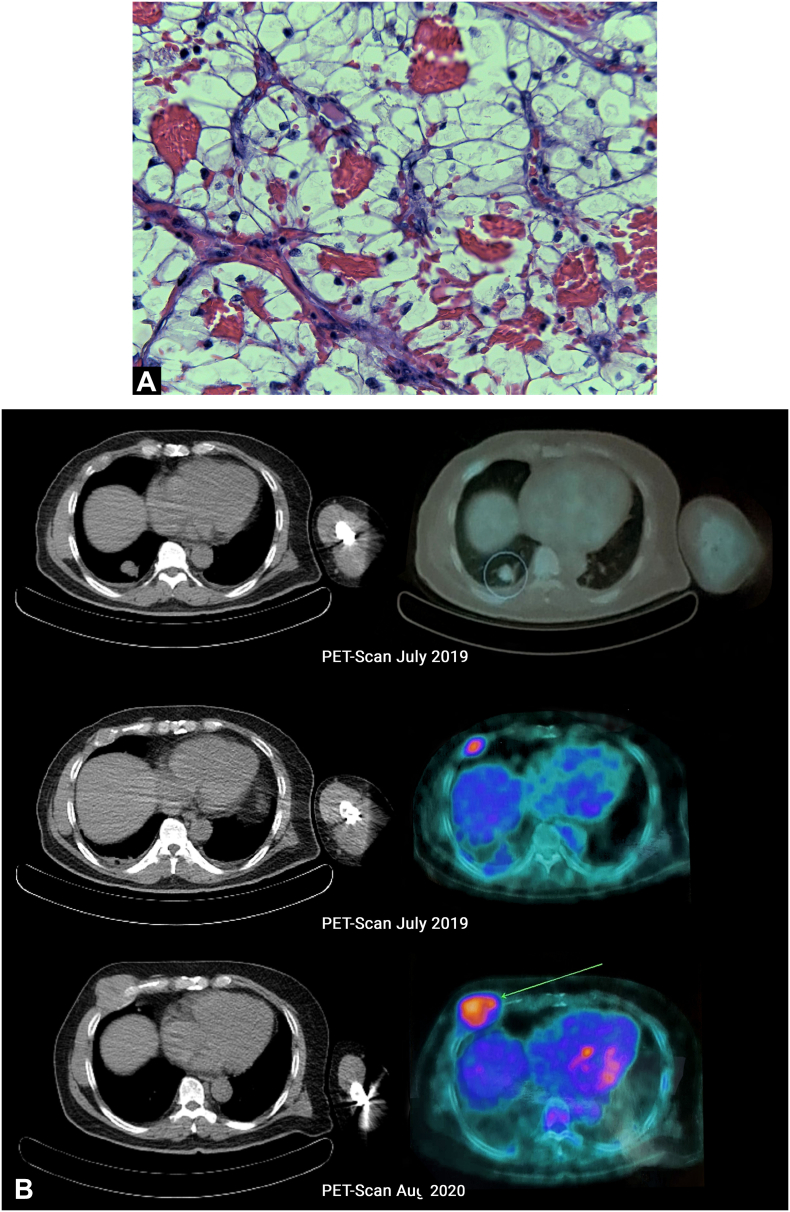
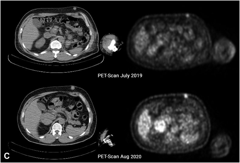
Case presentation: A 54-year-old male patient with renal cell carcinoma developed subcutaneous metastasis three months after radical nephrectomy and there was no discoloration or pain. Furthermore, an excision biopsy confirmed the metastatic lesion, and pazopanib was initiated as a treatment method. After 1-month of treatment, the patient developed ulceration and subsided after treatment was stopped. Similarly, a follow-up PET scan was performed almost a year after stopping the treatment, which showed improvement over metastatic pulmonary lesions.

Clinical discussion: Renal cell carcinoma (RCC) major metastases were observed in pulmonary, costal, and skin. Tumor burden and location of metastasis influences progression free-survival of RCC patients treated with TKI.

Conclusion: In this case, TKI treatment showed a long-term partial response, despite its lack of continuous therapy.

Keywords: Advanced cancer; Chemotherapy; Oncology; Renal cell carcinoma; Skin metastasis; Urology.


Conflict of interest statement

The authors had reported no conflicts of interest in this work.

Figures

Fig. 1
Fig. 1
(A) Left Renal Tumor on Coronal Plane CT-Scan with a size of 11.3 × 14.4 × 8.8 cm with contrast enhancement, no adrenal tissue could be identified suggestive that the tumor infiltrates the adrenal tissue. (B) Left Renal Tumor on Axial Planet CT-Scan, showing an inhomogen hipodense tumor occupied mostly on lateral side of the left kidney, expanding over the fascia gerota. (C) Confirmation of histological evaluation of left renal tumor infiltrates the adrenal tissue. (D) 40× magnification of the left renal tumor showed prominent nucleoli. Therefore, it was graded as WHO/ISUP grade 2.
Fig. 2
Fig. 2
(A) 40× magnification of left chest tumor showed prominent nucleoli and same characteristic that consist of solid cells with thin fibrovascular septa compared to the 40× magnification of the left renal tumor. (B) PET-Scan (July 2019) shows metabolic active multiple nodules on the 9th and 10th segment of the right lung and an active metabolic mass with a diameter of 2,6 cm on the 5th right anterior costae; On follow-up, PET-scan (August 2020) showed complete resolution of multiple nodules on the 9th and 10th segment of the right lung and progression of mass on the 5th right anterior costae to 5,7 cm (C) a mobile, firm, with a diameter of 1,3 cm metabolic active skin nodule is seen on the left upper abdominal and on follow-up PET-Scan its size increase to 1,8 cm.
Fig. 2
Fig. 2
(A) 40× magnification of left chest tumor showed prominent nucleoli and same characteristic that consist of solid cells with thin fibrovascular septa compared to the 40× magnification of the left renal tumor. (B) PET-Scan (July 2019) shows metabolic active multiple nodules on the 9th and 10th segment of the right lung and an active metabolic mass with a diameter of 2,6 cm on the 5th right anterior costae; On follow-up, PET-scan (August 2020) showed complete resolution of multiple nodules on the 9th and 10th segment of the right lung and progression of mass on the 5th right anterior costae to 5,7 cm (C) a mobile, firm, with a diameter of 1,3 cm metabolic active skin nodule is seen on the left upper abdominal and on follow-up PET-Scan its size increase to 1,8 cm.
Fig. 3
Fig. 3
(A) 1 month after ingestion of Pazopanib as systemic therapy, the patient began to develop ulceration over the surgical biopsy site on his left abdomen. (B) Four days after discontinuation of Pazopanib, wound improvement was shown. (C) Ulceration was almost completely resolved after eight days of Pazopanib discontinuation.

References

    1. Agha R.A., Franchi T., Sohrabi C., et al. The SCARE 2020 guideline: updating consensus Surgical CAse REport (SCARE) guidelines. Int. J. Surg. 2020;84:226–230. doi: 10.1016/j.ijsu.2020.10.034. - DOI - PubMed
    1. Wen L., Guo L., Zhang W., et al. Cooperation between the inflammation and coagulation systems promotes the survival of circulating tumor cells in renal cell carcinoma patients. Vol. 9. 2019. pp. 1–13. - DOI - PMC - PubMed
    1. Santoni M., Cimadamore A., Cheng L., et al. Circulating tumor cells in renal cell carcinoma: recent findings and future challenges. Front. Oncol. 2019;9:1–14. doi: 10.3389/fonc.2019.00228. - DOI - PMC - PubMed
    1. Bodnar R.J. Anti-angiogenic drugs: involvement in cutaneous side effects and wound-healing complication. Adv. Wound Care. 2014;3:635–646. doi: 10.1089/wound.2013.0496. - DOI - PMC - PubMed
    1. Czarnecka A.M., Brodziak A., Sobczuk P., et al. Metastatic tumor burden and loci as predictors of first line sunitinib treatment efficacy in patients with renal cell carcinoma. Sci. Rep. 2019;9:1–10. doi: 10.1038/s41598-019-44226-y. - DOI - PMC - PubMed