Review
. 2022 Mar 23;11(4):617.
doi: 10.3390/antiox11040617.

Contribution of Müller Cells in the Diabetic Retinopathy Development: Focus on Oxidative Stress and Inflammation

Affiliations
Review

Contribution of Müller Cells in the Diabetic Retinopathy Development: Focus on Oxidative Stress and Inflammation

Raul Carpi-Santos et al. Antioxidants (Basel). .

Abstract

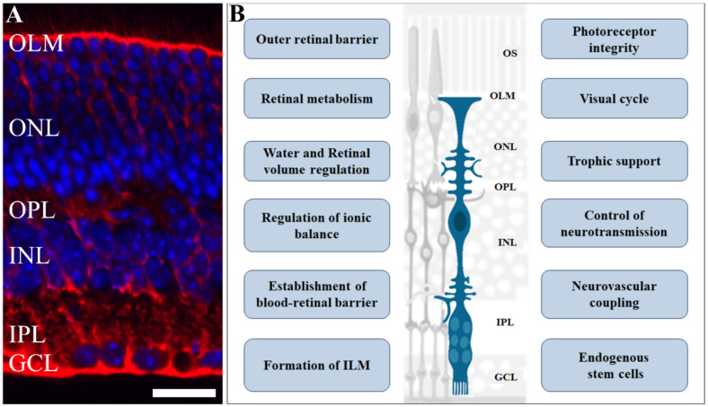
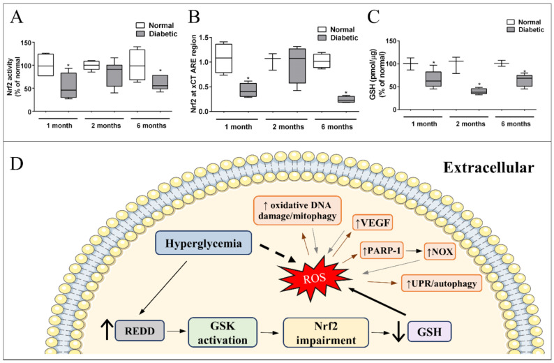
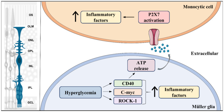
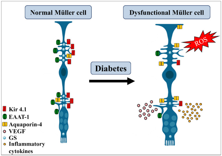
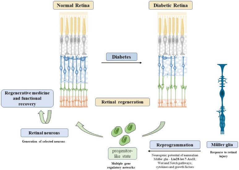
Diabetic retinopathy is a neurovascular complication of diabetes and the main cause of vision loss in adults. Glial cells have a key role in maintenance of central nervous system homeostasis. In the retina, the predominant element is the Müller cell, a specialized cell with radial morphology that spans all retinal layers and influences the function of the entire retinal circuitry. Müller cells provide metabolic support, regulation of extracellular composition, synaptic activity control, structural organization of the blood-retina barrier, antioxidant activity, and trophic support, among other roles. Therefore, impairments of Müller actions lead to retinal malfunctions. Accordingly, increasing evidence indicates that Müller cells are affected in diabetic retinopathy and may contribute to the severity of the disease. Here, we will survey recently described alterations in Müller cell functions and cellular events that contribute to diabetic retinopathy, especially related to oxidative stress and inflammation. This review sheds light on Müller cells as potential therapeutic targets of this disease.

Keywords: Müller glia; Nrf2; antioxidants; diabetes; reactive oxidative stress; retina.


Conflict of interest statement

The authors declare no conflict of interest.

Figures

Figure 1
Figure 1
Role of Müller glia in retinal physiology. Müller glia cells have an elaborate radial morphology, characterized by vertical processes, that span throughout the retina and branches in outer and inner layers: (A) Photomicrograph of retinal section immunolabelled for glutamine synthetase (red), a Müller cell marker, counterstained with DAPI (blue), showing the retinal cells nuclei. (B) A scheme highlighting various functions and events regulated by Müller cells in the retina such as retinal volume and ionic balance; blood–retinal barrier formation and maintenance; neurotransmission control, trophic support for other cells, and are an endogenous source of stem cells. Scale bar= 20 µm. OS: outer segment; OLM: outer limiting membrane; ONL: outer nuclear layer; OPL: outer plexiform layer; INL: inner plexiform layer; IPL: inner plexiform layer; GCL: ganglion cell layer. Adapted from smart.servier.com (accessed on 1 April 2021).
Figure 2
Figure 2
Signaling pathway underlying impaired Nrf2 function and effectors of excessive ROS in Müller cells in diabetes: (A) Nrf2 activity and (B) Binding to xCT ARE region, are decreased (↓) after 1 and 6 months of diabetes. (C) GSH is reduced in the retina of diabetic rats in early (1 month) and late periods (6 months). Modified from [237]. Copyright 2018 Elsevier. (D) Hyperglycemia induces an increase in REDD1 expression in Müller cells, which activates GSK. Once activated, GSK impairs Nrf2 function. As a result, there is a decrease in GSH levels and antioxidant defense capacity, resulting in higher oxidative stress in Müller glia. Direct effect of excessive ROS in Müller cells is indicated by brown arrows: increase (↑) in oxidative DNA damage/mitophagy, VEGF, poly [ADP-ribose] polymerase 1 (PARP-1), and unfolded protein response (UPR)/autophagy. All these responses intensify the production of ROS (gray arrows) enhancing oxidative stress in Müller cells. * p < 0.05 in comparison to normal.
Figure 3
Figure 3
Pro-inflammatory signal pathways activated in Müller glia in diabetes. Hyperglycemia leads to activation of multiple pro-inflammatory signaling pathways, such as Rock-1, c-myc, and CD40, which results in an initial increase (↑) in production of pro-inflammatory factors in Müller glia. In addition, CD40 activation triggers ATP release by Müller glia, which acts as an inflammatory inducer in monocytic cells through its receptor, P2X7. Left part of the figure was adapted from smart.servier.com (accessed on 1 April 2021).
Figure 4
Figure 4
Diabetes effects on Müller glia. Diabetic condition triggers several morphological and metabolic alterations in Müller glia cells. They become hypertrophic, suffer increase in aquaporin-4, decrease the expression of Kir4.1 channel, glutamine synthetase (GS), and EAAT-1 transporters, thus affecting the regulation of the ionic balance and glutamate uptake. Under this condition, Müller glia also increase the production of reactive oxidative species (ROS) and the release of pro-inflammatory cytokines and VEGF, contributing to retinal damage. EAAT, excitatory amino acid transporter. Adapted from smart.servier.com (accessed on 1 April 2021).
Figure 5
Figure 5
Reprogramming Müller cells in diabetic retinopathy. DR induces retinal neurodegeneration, an event difficult to recover. Alternatively, Müller reprogramming could act as a possible therapeutic option to restore visual capacity in DR patients. MGC reprogramming through activation of Lin28-let-7-Ascl1 genes, Wnt, and Notch pathways could lead to a progenitor-like state in these cells. Thus, these progenitors could be used to generate selected neurons, leading to retinal regeneration. Refs. [143,295,296,297] was adapted from smart.servier.com (accessed on 1 April 2021). Copyright 2016 Elsevier.

References

    1. Cho N.H., Shaw J.E., Karuranga S., Huang Y., da Rocha Fernandes J.D., Ohlrogge A.W., Malanda B. IDF Diabetes Atlas: Global Estimates of Diabetes Prevalence for 2017 and Projections for 2045. Diabetes Res. Clin. Pract. 2018;138:271–281. doi: 10.1016/j.diabres.2018.02.023. - DOI - PubMed
    1. Prevention of Diabetes Mellitus . Report of a WHO Study Group. Volume 844 WHO; Geneva, Switzerland: 1994. - PubMed
    1. Pereira-Figueiredo D., Brito R., Araújo D.S.M., Nascimento A.A., Lyra E.S.B., Cheibub A.M.S.S., Pereira Netto A.D., Ventura A.L.M., Paes-de-Carvalho R.C.K., Calaza K.C. Caffeine Exposure Ameliorates Acute Ischemic Cell Death in Avian Developing Retina. Purinergic Signal. 2020;16:41–59. doi: 10.1007/s11302-020-09687-1. - DOI - PMC - PubMed
    1. Lin A.D., Lee A.Y., Zhang Q., Rezaei K.A., Kinyoun J., Wang R.K., Lee C.S. Association Between OCT-based Microangiography Perfusion Indices and Diabetic Retinopathy Severity. Br. J. Ophthalmol. 2018;101:960–964. doi: 10.1136/bjophthalmol-2016-309514. - DOI - PMC - PubMed
    1. Hu L., Gong C., Chen X., Zhou H., Yan J., Hong W. Associations between Vascular Endothelial Growth Factor Gene Polymorphisms and Different Types of Diabetic Retinopathy Susceptibility: A Systematic Review and Meta-Analysis. J. Diabetes Res. 2021;2021:7059139. doi: 10.1155/2021/7059139. - DOI - PMC - PubMed

LinkOut - more resources