Review
. 2022 Mar 30;10(4):806.
doi: 10.3390/biomedicines10040806.

The Hallmarks of Glioblastoma: Heterogeneity, Intercellular Crosstalk and Molecular Signature of Invasiveness and Progression

Affiliations
Review

The Hallmarks of Glioblastoma: Heterogeneity, Intercellular Crosstalk and Molecular Signature of Invasiveness and Progression

Filippo Torrisi et al. Biomedicines. .

Abstract

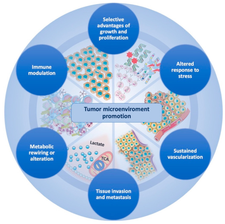
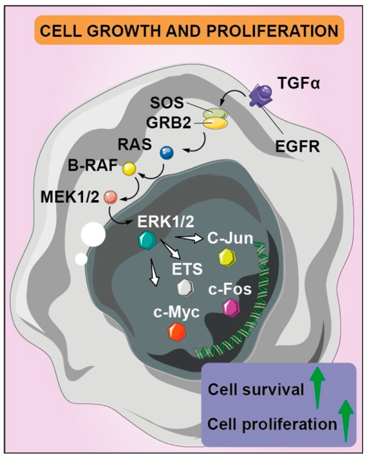
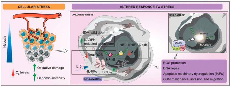
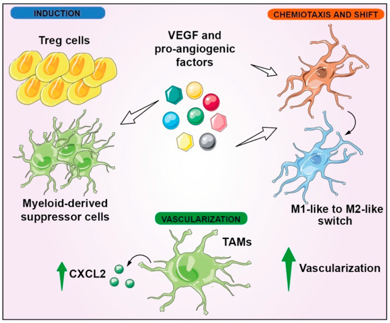
In 2021 the World Health Organization published the fifth and latest version of the Central Nervous System tumors classification, which incorporates and summarizes a long list of updates from the Consortium to Inform Molecular and Practical Approaches to CNS Tumor Taxonomy work. Among the adult-type diffuse gliomas, glioblastoma represents most primary brain tumors in the neuro-oncology practice of adults. Despite massive efforts in the field of neuro-oncology diagnostics to ensure a proper taxonomy, the identification of glioblastoma-tumor subtypes is not accompanied by personalized therapies, and no improvements in terms of overall survival have been achieved so far, confirming the existence of open and unresolved issues. The aim of this review is to illustrate and elucidate the state of art regarding the foremost biological and molecular mechanisms that guide the beginning and the progression of this cancer, showing the salient features of tumor hallmarks in glioblastoma. Pathophysiology processes are discussed on molecular and cellular levels, highlighting the critical overlaps that are involved into the creation of a complex tumor microenvironment. The description of glioblastoma hallmarks shows how tumoral processes can be linked together, finding their involvement within distinct areas that are engaged for cancer-malignancy establishment and maintenance. The evidence presented provides the promising view that glioblastoma represents interconnected hallmarks that may led to a better understanding of tumor pathophysiology, therefore driving the development of new therapeutic strategies and approaches.

Keywords: glioblastoma; immune modulation; invasiveness; metabolism; stress response.


Conflict of interest statement

The authors declare no conflict of interest.

Figures

Figure 1
Figure 1
Glioblastoma-tumor microenvironment promotion is ensured by the coexistence of interconnected cancer hallmarks that act with several strategies to achieve tumor maintenance and progression. The circular structure holding the hallmarks emphasizes the cooperation that GBM cells exert to create an ultimate tumor microenvironment that supports survival and progression of cancer cells.
Figure 2
Figure 2
Aberrant regulation of tyrosine kinase receptors determines several signaling pathways’ activation, including mitogen-activated protein kinase cascades with pleiotropic effects, such as cell proliferation and survival. EGFR and its mutated version EGFRvIII’s overexpression and/or hyper-activation is also fostered by autocrine feedback loop that leads to the transcription of EGFR ligands such as TGFα; the downstream effector Grb2 activates guanine nucleotide exchange factor SOS for GDP/GTP cycling, which promotes formation of active Ras-GTP, binding the other downstream effector targets such as B-Raf. It phosphorylates MEK1 and MEK2 dual-specificity protein kinases, which determine the ERK1 and ERK2 nuclear translocation, where they find several protein targets, such as transcription factors of Ets family, c-MYC and c-JUN. The latter forms a heterodimer with c-FOS, leading to increased cell survival, proliferation, and invasion.
Figure 3
Figure 3
Schematic representation of altered GBM cells responses to stress. The typical condition of hypoxia that characterize GBM TME leads to an increased amount of mitochondrial and intracellular ROS and heightened genome instability. Low levels of O2 induce stabilization and activation of HIF-1α/HIF-1β axis, which improves superoxide-anion (·O2) removal. Inflammatory state of hypoxic cells allows total ROS detoxification by IL-6/IL-6Rα enhancing and mitochondrial SOD-2 production, guided by NF-κB signaling-pathway activation. Moreover, restored levels of reduced glutathione by cytosolic and mitochondrial NADPH production in IDH wild-type GBM guarantee the protection from oxidative stress. Finally, stress condition also stimulates DDR, involving DNA-PK, ATR, and ATM recruitment, which ameliorates SSB/DSB repair system. All of these processes aim to overcome oxidative stress and DNA instability, ensuring increased GBM malignance, invasion, and migration.
Figure 4
Figure 4
Vascularization and immunomodulation interconnection in GBM. VEGF induces Treg cells and MDSCs; furthermore, it stimulates M1-like to M2-like switch in microglial cells, leading to immune evasion. Moreover, proangiogenic factors, including VEGF, induce chemiotaxis of microglial cells. Instead, resident TAMs promote vascularization, upregulating the cell-signaling cytokine CXCL2.
Figure 5
Figure 5
Tissue invasion and metastasis in GBM. Many pathways are involved in the invasion processes of GBM, especially in hypoxic and inflammatory contexts. Hypoxic and inflammatory conditions lead to the recruitment of myeloid cells, which promote TGF-β, EGF, PDGF, and FGF2 signaling, triggering the EMT initiation. In hypoxic conditions, HIFs regulate ZEB1, inhibiting E-cadherin expression, which contributes to the loss of cell–cell adhesion and the increase in motility, also supported by the activation of TWIST1, SRC, and chemokine receptors. In the inflammatory context, the upregulation of proinflammatory genes determines an increase in migration and proliferation, while ANX, through the FPR1 activation, induces the heightening of invasion and survival ability by means of STAT3, NF-kB, and HIF-1α pathways. Moreover, proliferation is also promoted by TH signaling, derived from microglia-GBM crosstalk.
Figure 6
Figure 6
Representation of metabolic reprogramming processes connected GBM invasiveness. The support of GBM invasiveness in hypoxic conditions is not only directly linked to lactate production, but also by indirect effects such as the production of additional signals of molecules such as PGI/AMF, and lipid metabolism and amino-acid reprogramming offers an important contribution to the proinvasion phenotype. Microglia adopts aerobic glycolysis by increasing lactate production and promoting M2 polarization with concomitant production of cytokines and other factors that can directly suppress effector cells, or indirectly via other types of immune cells such as intratumoral DCs and Treg cells, resulting in immunosuppression.
Figure 7
Figure 7
Representation of metabolic reprogramming processes connected GBM invasiveness. The support of GBM invasiveness in hypoxic conditions is not only directly linked to lactate production, but also by indirect effects such as the production of additional signals of molecules such as phosphoglucose isomerase/autocrine-motility factor (PGI/AMF), lipid metabolism and amino-acid reprogramming offer an important contribution to the proinvasion phenotype. Microglia adopts aerobic glycolysis by increasing lactate production and promoting M2 polarization with the concomitant production of cytokines and other factors that can directly suppress effector cells or indirectly via other types of immune cells such as intratumoral DCs and Treg cells, resulting in immunosuppression.

References

    1. Jeibouei S., Akbari M.E., Kalbasi A., Aref A.R., Ajoudanian M., Rezvani A., Zali H. Personalized medicine in breast cancer: Pharmacogenomics approaches. Pharm. Pers. Med. 2019;12:59–73. doi: 10.2147/PGPM.S167886. - DOI - PMC - PubMed
    1. Prat A., Parker J.S., Karginova O., Fan C., Livasy C., Herschkowitz J.I., He X., Perou C.M. Phenotypic and molecular characterization of the claudin-low intrinsic subtype of breast cancer. Breast Cancer Res. 2010;12:R68. doi: 10.1186/bcr2635. - DOI - PMC - PubMed
    1. Chan C.W.H., Law B.M.H., So W.K.W., Chow K.M., Waye M.M.Y. Novel Strategies on Personalized Medicine for Breast Cancer Treatment: An Update. Int. J. Mol. Sci. 2017;18:2423. doi: 10.3390/ijms18112423. - DOI - PMC - PubMed
    1. Yersal O., Barutca S. Biological subtypes of breast cancer: Prognostic and therapeutic implications. World J. Clin. Oncol. 2014;5:412–424. doi: 10.5306/wjco.v5.i3.412. - DOI - PMC - PubMed
    1. Ostrom Q.T., Cioffi G., Gittleman H., Patil N., Waite K., Kruchko C., Barnholtz-Sloan J.S. CBTRUS Statistical Report: Primary Brain and Other Central Nervous System Tumors Diagnosed in the United States in 2012–2016. Neuro-Oncology. 2019;21:v1–v100. doi: 10.1093/neuonc/noz150. - DOI - PMC - PubMed

LinkOut - more resources