. 2022 Apr 6;10(4):856.
doi: 10.3390/biomedicines10040856.

The Impact of Prolonged Inflammation on Wound Healing

Affiliations

The Impact of Prolonged Inflammation on Wound Healing

Judith C J Holzer-Geissler et al. Biomedicines. .

Abstract

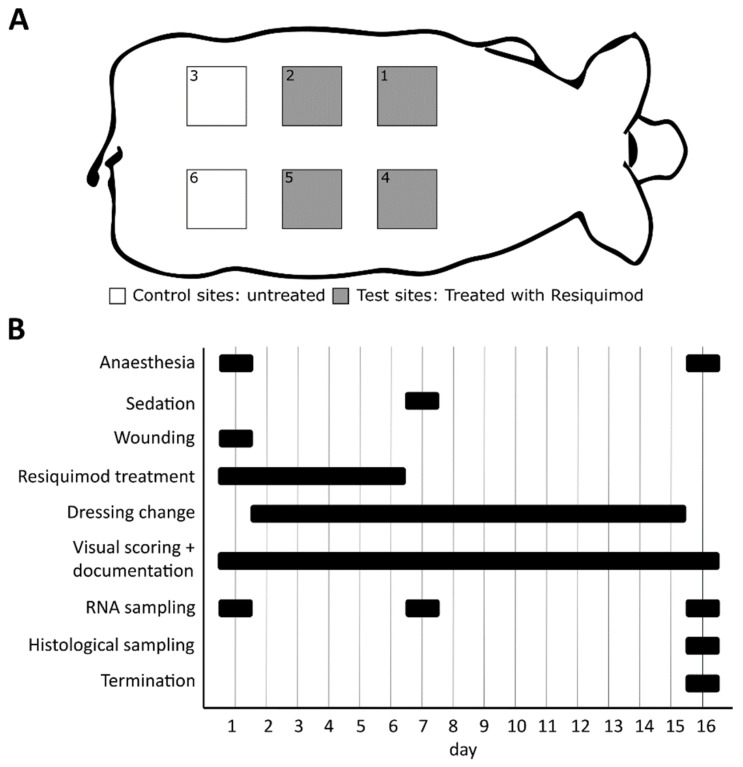
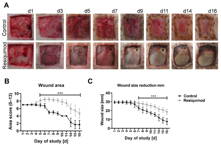
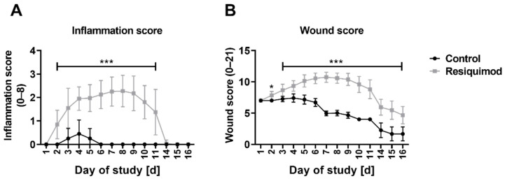
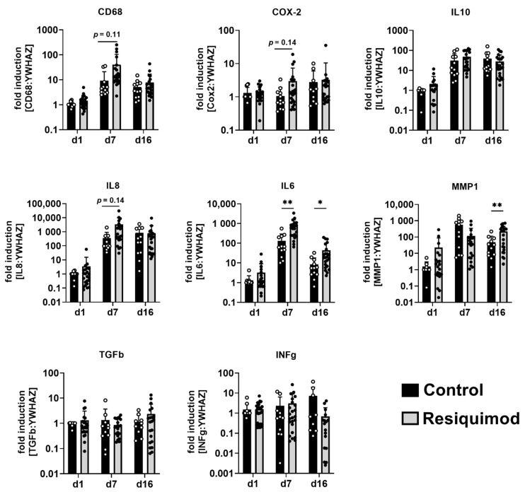
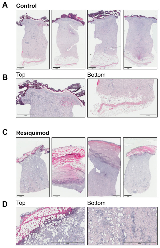
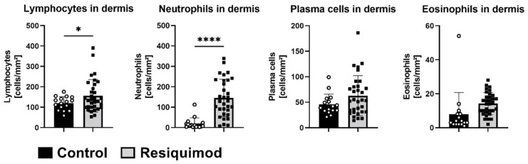
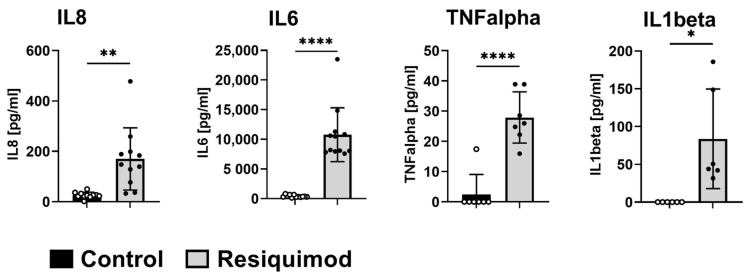
The treatment of chronic wounds still challenges modern medicine because of these wounds' heterogenic pathophysiology. Processes such as inflammation, ischemia and bacterial infection play major roles in the progression of a chronic wound. In recent years, preclinical wound models have been used to understand the underlying processes of chronic wound formation. However, the wound models used to investigate chronic wounds often lack translatability from preclinical models to patients, and often do not take exaggerated inflammation into consideration. Therefore, we aimed to investigate prolonged inflammation in a porcine wound model by using resiquimod, a TLR7 and TLR8 agonist. Pigs received full thickness excisional wounds, where resiquimod was applied daily for 6 days, and untreated wounds served as controls. Dressing change, visual documentation and wound scoring were performed daily. Biopsies were collected for histological as well as gene expression analysis. Resiquimod application on full thickness wounds induced a visible inflammation of wounds, resulting in delayed wound healing compared to non-treated control wounds. Gene expression analysis revealed high levels of IL6, MMP1 and CD68 expression after resiquimod application, and histological analysis showed increased immune cell infiltration. By using resiquimod, we were able to show that prolonged inflammation delayed wound healing, which is often observed in chronic wounds in patients. The model we used shows the importance of inflammation in wound healing and gives an insight into the progression of chronic wounds.

Keywords: inflammation; prolonged inflammation; resiquimod R848; wound healing; wound model.


Conflict of interest statement

The authors declare no conflict of interest.

Figures

Figure 1
Figure 1
Schematic presentation of the experiment setup. (A) Each pig (n = 10) received 6 wounds on its back, 4 of which were treated with resiquimod, and 2 of which remained untreated as control wounds. Control and resiquimod-induced wounds were randomly arranged on the back of each pig to avoid a location bias. (B) The entire duration of the experiment was 16 days, during which the animals were anesthetised twice and received a sedation once. Resiquimod application lasted for 6 days. Dressing changes were performed on a daily basis, as were visual scoring and documentation. Biopsies for gene expression analysis were collected on days 1, 7 and 16, and biopsies for histology were collected only on day 16.
Figure 2
Figure 2
Wound morphology and wound area over the course of the experiment. Wounds of 3 × 3 cm were inflicted on the backs of pigs. Resiquimod was applied for 6 days, and the wounds were further examined over the course of 10 days. (A) Daily visual documentation and scoring was done. The control wounds ((A)–top row) showed physiological wound healing as well as wound contracture. The resiquimod-induced wounds ((A)–bottom row) showed a delay in wound healing and the formation of necrotic scabs. (B) The area scores showed higher values in resiquimod-induced wounds. (C) The wound size was determined by measuring the width of the wound, and less wound size reduction was shown in resiquimod-induced wounds. Data are presented as mean (dot) ± SD (whiskers) (n = 20 for control wounds; n = 40 for resiquimod-induced wounds). Data shown as mean ± SD. Statistical significance has been determined by 2-way ANOVA and corrected for multiple comparisons using the Sidak method. *** (p < 0.001).
Figure 3
Figure 3
Inflammation score and wound score over the course of the experiment. (A) The inflammation score showed a highly significant difference between control and resiquimod-induced wounds. (B) The overall wound score includes the area score as well as the inflammation score. Data are presented as mean (dot) ± standard deviation (whiskers) (n = 20 for control wounds; n = 40 for resiquimod-induced wounds). Data shown as mean ± SD. Statistical significance has been determined by 2-way ANOVA and corrected for multiple comparisons using the Sidak method. *** (p < 0.001).
Figure 4
Figure 4
Resiquimod-induced inflammation increased the expression of IL6, MMP1, CD68, IL8 and COX-2. Relative mRNA expression levels of CD68, COX-2, IL10, IL8, IL6, MMP1, TGFb and INFg were determined by qPCR, normalising target gene expression to the averaged expression of YWHAZ. Resiquimod-induced wounds (grey) were compared to control wounds (black). Data are presented as mean (bar) + standard deviation (whiskers) (n = 9–12 for controls and 17–24 for resiquimod application). Data shown as mean + SD. Statistical significance has been determined by 2-way ANOVA and corrected for multiple comparisons using the Sidak method. * (p < 0.05), ** (p < 0.01).
Figure 5
Figure 5
Resiquimod-induced inflammation leads to increased immune cell infiltration. (A) Four representative full overviews of haematoxylin/eosin staining of control wounds. (B) Detailed overview of one representative picture (left-top of the section); stratum corneum, epidermis and dermis of control wounds (right-bottom of the section) and dermis and subcutaneous adipose tissue of control wounds. Control wounds show physiological layers of skin, including a scab on top of the epidermis and a physiological layer of subcutaneous adipose tissue. (C) Four representative full overviews of haematoxylin/eosin staining of resiquimod-induced wounds. (D) Detailed overview of one representative picture (left-top of the section) of a barely existing stratum corneum, fully altered epidermis and dermis of resiquimod-induced wounds and (right-bottom of the section) a dermis with dispersed adipocytes in resiquimod-induced wounds. Resiquimod-induced wounds show high immune cell infiltration and no visible skin layers except the dermis. Scale bar = 1 mm.
Figure 6
Figure 6
Resiquimod-induced inflammation increases lymphocyte and neutrophil infiltration. Immune cells were counted in haematoxylin- and eosin-stained sections from biopsies at the study’s end (day 16). Lymphocytes and neutrophils were significantly increased in the dermises of resiquimod-induced wounds, whereas no differences in plasma cell or eosinophil counts were detected (n = 16 for control wounds and 32 for resiquimod-induced wounds). Data shown as mean ± SD. Statistical significance has been determined by unpaired t-test with a Welch’s correction. * (p < 0.05), **** (p < 0.0001).
Figure 7
Figure 7
Short-term resiquimod application promotes the release of cytokines in the dermis. Release of cytokines into dISF was analysed by OFM. Split-thickness wounds were inflicted and treated with resiquimod and, 48 h later, OFM probes were implanted to sample dISF. Total IL8, IL6, TNFalpha and IL1b content was measured (n = 6–12 per treatment). Data are shown as mean ± SD. Statistical significance was determined by unpaired t-test with a Welch’s correction. * (p < 0.05), ** (p < 0.01), **** (p < 0.0001).

References

    1. Lazarus G., Valle M.F., Malas M., Qazi U., Maruthur N.M., Doggett D., Fawole O.A., Bass E.B., Zenilman J. Chronic venous leg ulcer treatment: Future research needs. Wound Repair Regen. 2014;22:34–42. doi: 10.1111/wrr.12102. - DOI - PubMed
    1. Olsson M., Järbrink K., Divakar U., Bajpai R., Upton Z., Schmidtchen A., Car J. The humanistic and economic burden of chronic wounds: A systematic review. Wound Repair Regen. 2019;27:114–125. doi: 10.1111/wrr.12683. - DOI - PubMed
    1. Velnar T., Bailey T., Smrkolj V. The wound healing process: An overview of the cellular and molecular mechanisms. J. Int. Med. Res. 2009;37:1528–1542. doi: 10.1177/147323000903700531. - DOI - PubMed
    1. Hunt T.K., Hopf H., Hussain Z. Physiology of Wound Healing. Adv. Ski. Wound Care. 2000;13:1–13. doi: 10.1016/S0196-0644(88)80351-2. (Adv Skin Wound Care) - DOI - PubMed
    1. Ganesh K., Sinha M., Mathew-Steiner S.S., Das A.K., Roy S., Sen C.K. Chronic Wound Biofilm Model. Adv. Wound Care. 2015;4:382–388. doi: 10.1089/wound.2014.0587. - DOI - PMC - PubMed

LinkOut - more resources