. 2022 Apr 6;10(4):859.
doi: 10.3390/biomedicines10040859.

A Small Molecule Promoting Neural Differentiation Suppresses Cancer Stem Cells in Colorectal Cancer

Affiliations

A Small Molecule Promoting Neural Differentiation Suppresses Cancer Stem Cells in Colorectal Cancer

Jung Kyu Choi et al. Biomedicines. .

Abstract

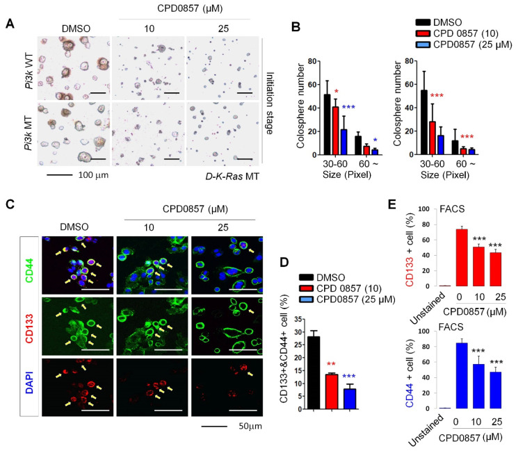
Cancer stem cells (CSCs) are a tumor cell subpopulation that drives tumor progression and metastasis, leading to a poor overall survival of patients. In colorectal cancer (CRC), the hyper-activation of Wnt/β-catenin signaling by a mutation of both adenomatous polyposis coli (APC) and K-Ras increases the size of the CSC population. We previously showed that CPD0857 inactivates Wnt/β-catenin signaling by promoting the ubiquitin-dependent proteasomal degradation of β-catenin and Ras proteins, thereby decreasing proliferation and increasing the apoptosis of CRC lines. CPD0857 also decreased the growth and invasiveness of CRC cells harboring mutant K-Ras resistant to EGFR mAb therapy. Here, we show that CPD0857 treatment decreases proliferation and increases the neuronal differentiation of neural progenitor cells (NPCs). CDP0857 effectively reduced the expression of CSC markers and suppressed self-renewal capacity. CPD0857 treatment also inhibited the proliferation and expression of CSC markers in D-K-Ras MT cells carrying K-Ras, APC and PI3K mutations, indicating the inhibition of PI3K/AKT signaling. Moreover, CPD0857-treated xenograft mice showed a regression of tumor growth and decreased numbers of CSCs in tumors. We conclude that CPD0857 could serve as the basis of a drug development strategy targeting CSCs activated through Wnt/β-catenin-Ras MAPK-PI3K/AKT signaling in CRCs.

Keywords: K-Ras; Wnt/β-catenin; cancer stem cell; colorectal cancer; neural progenitor cell.


Conflict of interest statement

The authors declare no competing interests.

Figures

Figure 1
Figure 1
Screening drugs promoting neuronal differentiation of the NPCs via inhibiting Wnt/β-catenin signaling. (A) Nine lead compounds selected by a dual high-throughput screening system, which was described previously [32], were tested for measuring neuronal differentiation using primary NPCs derived from the E14.5 rat embryo forebrain. NPCs were treated with 9 compounds (10 μM) for 48 h in the presence or absence of 20 ng/ml bFGF. Micrographs were taken at a magnification of 200×. Scale bars: 50 μm. (B) Neurite outgrowths were quantified using ImageJ software. All data are shown as mean ± SD for at least 3 independent images. *** p  < 0.005, 2-sided t test. (C) NPCs were grown for 18 h in N2:B27 (1:1) medium containing bFGF and treated with VPA (negative control, 1mM) and each compound (10 μM). WCLs were immunoblotted using indicated antibodies. (D) Relative band intensity shown in (C) were quantified using ImageJ software (n = 3). Red arrows indicate significant decreases or increases in PCNA or Tuj1 in NPCs, respectively. (E) NPCs were transfected with Topflash reporter plasmid and co-transfected with β-galactosidase. Then, 24 h later, the growth medium containing transfection reagents was removed and replaced with N2 media with or without 25 μM CDP0857 for a further 24 h. Cells were then harvested and luciferase reporter assays were performed. (F) Western blot (WB) analysis of NPCs using indicated antibodies. (G) Relative band intensities shown in (F) were quantified using ImageJ software (n > 2). (H) Ubiquitylation assay of β-catenin and Ras proteins in indicated lysates of NPCs. Cells were transduced with HA-Ub plasmids and then treated a day later with or without CPD0857 for 24 h. Cells were also treated with MG132 (10 μg/mL) during 6 h before harvest. Immunoprecipitation (IP) was performed with β-catenin or Ras antibodies. WCLs were analyzed by WB for indicated antibodies (n = 2). (I,J) Relative band intensity of ubiquitylated proteins shown in (H) were quantified using ImageJ software (n = 3). Values correspond to mean ± SD. ANOVA tests were performed to calculate significance (* p < 0.05, ** p < 0.005, *** p < 0.005). NPCs, neural progenitor cells; VPA, valproic acid; WCLs, whole cell lysates; Ub, ubiquitin.
Figure 2
Figure 2
The effect of CDP0857 on proliferation and differentiation of the NPCs. (A) NPCs were grown in medium in the presence or absence of bFGF (20 ng/mL) and were treated with AG1478 (EGFR tyrosin kinase inhibitor, 20 μM), Cetuximab (EGFR monoclonal antibody, 10 μM), 5-FU (antineoplastic drug, 20 ug/mL), LY294002 (PI3K/AKT inhibitor, 20 μM), and CDP0857 (25 μM) during 24 h. Cells were subjected to immunofluorescent labeling using antibody specific for Nestin. Nuclei were counterstained with DAPI. Images were obtained using a Zeiss confocal microscope. Scale bars: 50 μm. (B) Quantification of Ki67/DAPI cells in analysis shown in (A, right) (n = 3). (C) The intensity of Nestin (blue) was quantified by using ImageJ software (n > 3). (D) NPCs were treated with CDP0857 (25 μM) during 48 h. Immunostaining with Nestin (red) antibody. DAPI (blue). Images were obtained using a Zeiss confocal microscope. (E) Quantification of Nestin/DAPI cells in analysis shown at (D) (n = 3). (F) RT-PCR analysis of indicated mRNAs in analysis shown in (D). (G) NPCs were treated with CDP0857 (25 μM) during 48 h. Proliferation and neuronal differentiation was monitored by BrdU incorporation and Tuj1 staining using immunocytochemical analysis. Nuclei were counterstained with DAPI. Scale bars: 50 μm. (H,I) Percentages of relative numbers of BrdU-positive cells (H) and Tuj1-positive cells (I). Values correspond to mean ± SD. ANOVA tests were performed to calculate significance (* p < 0.05, *** p < 0.0005). bFGF, basic fibroblast growth factor; BrdU, 5-bromo-2-deoxyuridine; DAPI, 40,6-diamidino-2-phenylindole.
Figure 3
Figure 3
Effects of CDP0857 on sphere formation and cancer stem cell activation of colorectal cancer (CRC) cells carrying K-Ras, APC and PI3K mutations. (AC) D-K-Ras WT and D-K-Ras MT cells harboring mutations of both APC and PI3K were seeded at a density of 2 × 10⁴ (n = 3) cells/plate in ultra-low-attachment 6-well plates for 5 days, and were then treated with varying doses of CDP0857 for 72 h. Scale bars, 100 μm. (B) Relative numbers and sizes of colospheres were measured using ImageJ software. (C) D-K-Ras MT cells harboring mutation both APC and PI3K were seeded at a density of 2 × 10⁴ cells/well in 24-well plates and were treated with varying doses of CDP0857 for 24 h. Immunofluorescent labeling of CD133 and CD44. Nuclei were counterstained with DAPI. Images were obtained using a Zeiss confocal microscope. Scale bars, 50 μm. (D) Quantification of CD133 and CD44-double-positive populations in each immunostained tumor cell. (E) The relative numbers of CD133- or CD44-positive cells were quantified by fluorescent-activated cell sorter (FACS) analyses. WT, wild-type; MT, mutant-type. * p < 0.05, ** p < 0.005, *** p < 0.005.
Figure 4
Figure 4
CDP0857 inhibits progression of tumor with mutations of K-Ras, APC and PI3K by reducing cancer stem cell populations. (A) Colon cancer cell lines were subjected to immunofluorescence analysis with indicated antibodies, and CD133- and CD44-double-positive populations in each immunstained tumor cell were quantified. Nuclei were counter-stained with DAPI. Images were captured using a Zeiss confocal microscope. Scale bars: 25 μm. (B) D-K-Ras MT cells harboring mutation both APC and PI3K were subcutaneously injected into flanks of BALB/c nude mice (n = 4). Mice were treated for 21 days by intra peritoneal (I.P.) injection with vehicle or with 25 mg/kg CDP0857 once every three days. Gross images showing representative tumor cases treated with drugs, with enlarged tumor images for each case presented at the bottom. (C) Tumor volumes of mice treated with vehicle or CDP0857 were measured with the Vernier calipers every 3 days. (D) Paraformaldehyde (PFA)-fixed paraffin sections from excised tumors were incubated with antibodies against β-catenin, pan-Ras, pERK, PCNA, CD133, CD44, and EpCAM. Images were captured using a Zeiss confocal microscope. Representative images were selected from at least 3 different fields. Scale bars, 50 μm. (E) Expression of antibodies in the immunostained tumor tissues in each representative xenograft tumor was quantified using ImageJ software. All data are shown as mean ± SD for at least 3 independent specimens. ** p  < 0.005, *** p  < 0.0005, 2-sided t test.

Similar articles

Cited by

References

    1. Siegel R.L., Miller K.D., Jemal A. Cancer statistics. CA Cancer J. Clin. 2018;68:7–30. doi: 10.3322/caac.21442. - DOI - PubMed
    1. Barker N., Ridgway R.A., van Es J.H., van de Wetering M., Begthel H., van den Born M., Danenber E., Clarke A.E., Sanson O.J., Clevers H. Crypt stem cells as the cells-of-origin of intestinal cancer. Nature. 2009;457:608–611. doi: 10.1038/nature07602. - DOI - PubMed
    1. Merlos-Suarez A., Barriga F.M., Jung P., Iglesias M., Cespedes M.V., Rossell D., Sevillano M., Hernando-Momblona X., da Silva-Diz V., Muñoz P., et al. The intestinal stem cell signature identifies colorectal cancer stem cells and predicts disease relapse. Cell Stem Cell. 2011;8:511–524. doi: 10.1016/j.stem.2011.02.020. - DOI - PubMed
    1. Zeuner A., De Maria R. Not so lonely at the top for cancer stem cells. Cell Stem Cell. 2011;9:289–290. doi: 10.1016/j.stem.2011.09.006. - DOI - PubMed
    1. Ro E.J., Cho Y.H., Jeong W.J., Park J.C., Min D.S., Choi K.-Y. WDR76 degrades RAS and suppresses cancer stem cell activation in colorectal cancer. Cell Commun. Signal. 2019;17:88. doi: 10.1186/s12964-019-0403-x. - DOI - PMC - PubMed