Review
. 2022 Apr 7;10(4):864.
doi: 10.3390/biomedicines10040864.

Group I mGluRs in Therapy and Diagnosis of Parkinson's Disease: Focus on mGluR5 Subtype

Affiliations
Review

Group I mGluRs in Therapy and Diagnosis of Parkinson's Disease: Focus on mGluR5 Subtype

Shofiul Azam et al. Biomedicines. .

Abstract

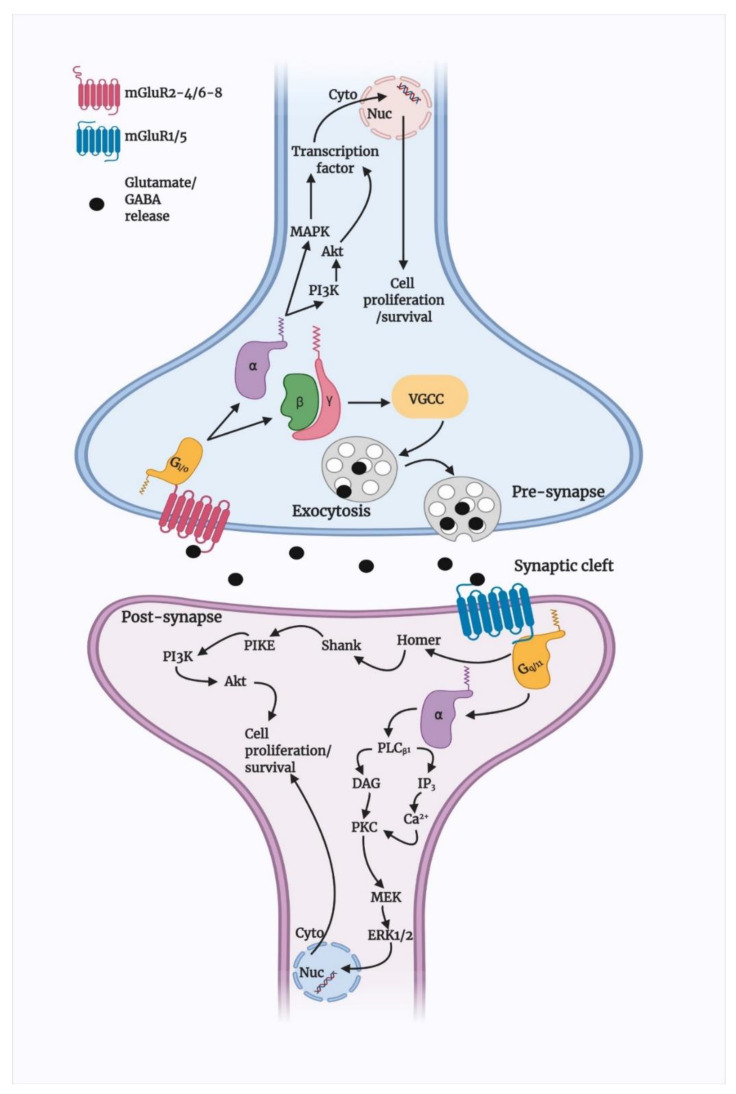
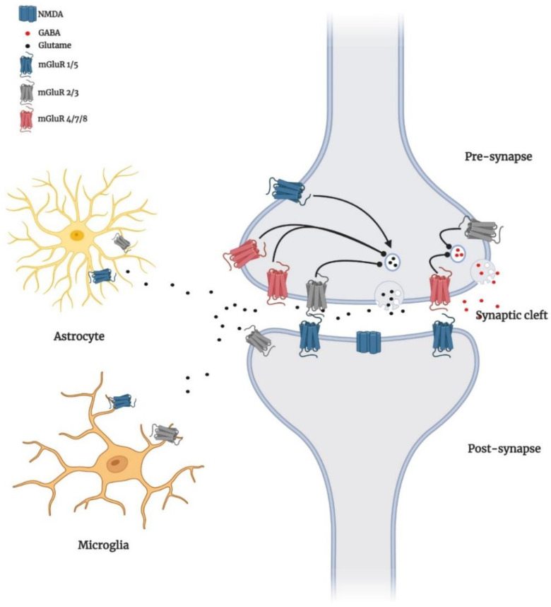
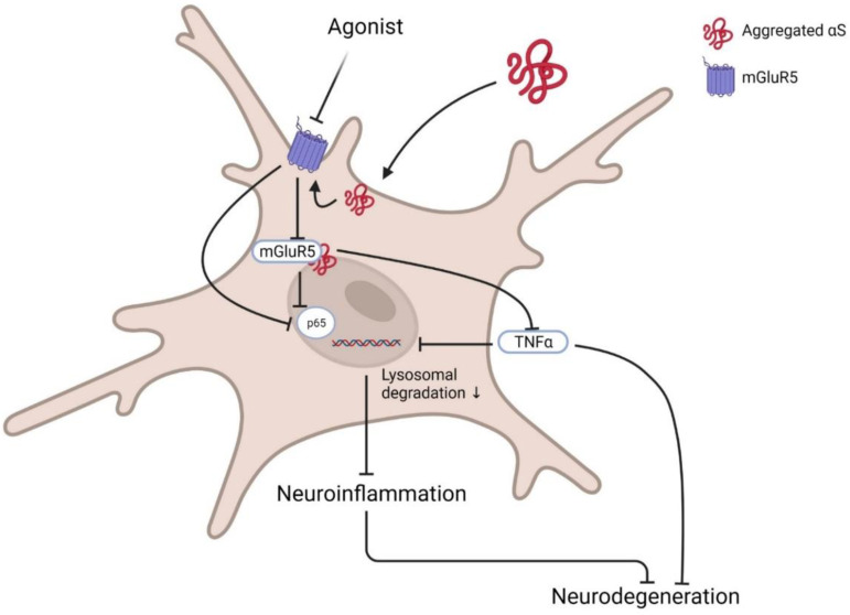
Metabotropic glutamate receptors (mGluRs; members of class C G-protein-coupled receptors) have been shown to modulate excitatory neurotransmission, regulate presynaptic extracellular glutamate levels, and modulate postsynaptic ion channels on dendritic spines. mGluRs were found to activate myriad signalling pathways to regulate synapse formation, long-term potentiation, autophagy, apoptosis, necroptosis, and pro-inflammatory cytokines release. A notorious expression pattern of mGluRs has been evident in several neurodegenerative diseases, including Alzheimer's disease, Parkinson's disease, Huntington's disease, and schizophrenia. Among the several mGluRs, mGluR5 is one of the most investigated types of considered prospective therapeutic targets and potential diagnostic tools in neurodegenerative diseases and neuropsychiatric disorders. Recent research showed mGluR5 radioligands could be a potential tool to assess neurodegenerative disease progression and trace respective drugs' kinetic properties. This article provides insight into the group I mGluRs, specifically mGluR5, in the progression and possible therapy for PD.

Keywords: C G-protein-coupled receptors; glutamate signalling; metabotropic glutamate receptors; neurodegenerative diseases; positron emission tomography; radioligands.


Conflict of interest statement

The authors declare no conflict of interest.

Figures

Figure 1
Figure 1
Schematic presentation of mGluRs cell signalling pathways. Likely, other GPCRs and mGluRs are located at the cell membrane that binds to extracellular substances and transmits signals to intracellular molecules called G-protein (G-α, -β, and -γ) Upon agonist activation, both group II and III mGluRs are coupled predominantly to Gi/o proteins, which mediate the downstream inhibition of adenylyl cyclase activity via Gαi/o, decreasing the levels of cAMP. Gβγ subunits modulate voltage-dependent ion channels, inhibiting Ca2+ and limiting presynaptic glutamate or GABA release. Group II and III mGluRs also activate PI3K/Akt and MAPK pathways and enhance neuroprotection by increasing the production of neurotrophic factors [58]. In the post-synapse, following glutamate binding, group I mGluRs uncouple Gαq/11 proteins, stimulate PLCβ1, and activate DAG and IP3, which increase intracellular Ca2+ efflux. Both Ca2+ and DAG activate PKC, which has been proposed to activate MEK/ERK1/2 signalling. Furthermore, mGluR1/5 interacts with Homer proteins, which activate Shank proteins. This complex of Homer proteins and group I mGluRs activates Akt through a mechanism that involves PI3K, PDK1, and PIKE, promoting neuroprotection [8]. Physical interaction between elements is represented by a continuous line.
Figure 2
Figure 2
Schematic display of the distribution of mGluRs. Group I mGluRs (blue) are localised post-synaptically, and group II (gray) and III (red) receptors are localised in presynaptic neurons. However, exceptions occur; for example, mGluR5 and mGluR3 are widely expressed in glial cells (astrocyte and microglial cells) [54], although signalling and consequences of mGluR in glial cells have yet to be fully uncovered but are now considered as an emerging key site of synaptic mGluR regulation. In presynaptic locations, mGluRs 2, 3, 4, 7, and 8 are generally found in extrasynaptic areas and inhibit the release of glutamate (black circles) or GABA (red circles). In contrast, group I receptors promote release when present. At the postsynaptic terminal, the glutamate-gated ion channel NMDA responds to glutamate with increases in intracellular sodium or calcium, promoting cell excitability. mGluR5 and NMDA receptors are closely linked signalling partners reciprocally regulated by phosphorylation. Postsynaptic mGluR2/3 receptors couple to cAMP inhibition.
Figure 3
Figure 3
Basic pathway of mGluR5 agonist in PD. Post activation of microglia by the presence of αS at the membrane inactivates membrane receptor mGluR5 and its downstream G-protein. Sequentially activated transcription factors NFκB p65 and TNFα enhance inflammatory cytokine release and induce neurodegeneration. Agonists such as CPHG could ameliorate this pathway by increasing mGluR5 expression via reducing lysosomal degradation, further downregulating related inflammatory signalling activation and subsequent inhibition of microglia-mediated inflammation to prevent neurotoxicity. Created with BioRender.com (accessed on 31 March 2022).

References

    1. Ferraguti F., Crepaldi L., Nicoletti F. Metabotropic Glutamate 1 Receptor: Current Concepts and Perspectives. Pharmacol. Rev. 2008;60:536–581. doi: 10.1124/pr.108.000166. - DOI - PubMed
    1. Jakaria M., Park S.-Y., Haque M.E., Karthivashan G., Kim I.-S., Ganesan P., Choi D.-K. Neurotoxic Agent-Induced Injury in Neurodegenerative Disease Model: Focus on Involvement of Glutamate Receptors. Front. Mol. Neurosci. 2018;11:307. doi: 10.3389/fnmol.2018.00307. - DOI - PMC - PubMed
    1. Dingledine R., Borges K., Bowie D., Traynelis S.F. The glutamate receptor ion channels. Pharmacol. Rev. 1999;51:7–61. - PubMed
    1. Pin J.-P., Galvez T., Prézeau L. Evolution, structure, and activation mechanism of family 3/C G-protein-coupled receptors. Pharmacol. Ther. 2003;98:325–354. doi: 10.1016/S0163-7258(03)00038-X. - DOI - PubMed
    1. Willard S.S., Koochekpour S. Glutamate, Glutamate Receptors, and Downstream Signaling Pathways. Int. J. Biol. Sci. 2013;9:948–959. doi: 10.7150/ijbs.6426. - DOI - PMC - PubMed

LinkOut - more resources