. 2022 Apr 9;10(4):873.
doi: 10.3390/biomedicines10040873.

Proteasome Inhibitors Decrease the Viability of Pulmonary Arterial Smooth Muscle Cells by Restoring Mitofusin-2 Expression under Hypoxic Conditions

Affiliations

Proteasome Inhibitors Decrease the Viability of Pulmonary Arterial Smooth Muscle Cells by Restoring Mitofusin-2 Expression under Hypoxic Conditions

I-Chen Chen et al. Biomedicines. .

Abstract

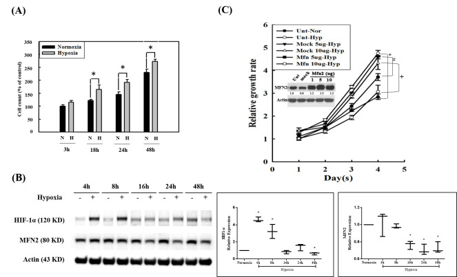
Pulmonary hypertension (PH) is a severe progressive disease, and the uncontrolled proliferation of pulmonary artery smooth muscle cells (PASMCs) is one of the main causes. Mitofusin-2 (MFN2) profoundly inhibits cell growth and proliferation in a variety of tumor cell lines and rat vascular smooth muscle cells. Down-regulation of MFN2 is known to contribute to PH. Proteasome inhibitors have been shown to inhibit the proliferation of PASMCs; however, there is no study on the regulation of proteasome inhibitors through MFN-2 in the proliferation of PASMCs, a main pathophysiology of PH. In this study, PASMCs were exposed to hypoxic conditions and the expression of MFN2 and cleaved-PARP1 were detected by Western blotting. The effects of hypoxia and proteasome inhibitors on the cell viability of PASMC cells were detected by CCK8 assay. The results indicated that hypoxia increases the viability and reduces the expression of MFN2 in a PASMCs model. MFN2 overexpression inhibits the hypoxia-induced proliferation of PASMCs. In addition, proteasome inhibitors, bortezomib and marizomib, restored the decreased expression of MFN2 under hypoxic conditions, inhibited hypoxia-induced proliferation and induced the expression of cleaved-PARP1. These results suggest that bortezomib and marizomib have the potential to improve the hypoxia-induced proliferation of PASMCs by restoring MFN2 expression.

Keywords: hypoxia; mitofusin-2; proteasome inhibitor; pulmonary hypertension.


Conflict of interest statement

The authors declare no conflict of interest.

Figures

Figure 1
Figure 1
The role of MFN2 in hypoxia-induced proliferation. (A) HPASMCs were exposed to hypoxic (1% oxygen) or normoxic conditions for 3, 18, 24, and 48 h, respectively. Cell viability was detected using a CCK8 assay. * p < 0.05 hypoxic group versus normoxic group. The statistical difference was analyzed by an unpaired t-test and the data represented by means ±standard error of three independent experiments. (B) HPASMCs were exposed to the hypoxic (+) or normoxic (−) conditions for different times. Expression of HIF-1α and MFN2 was detected by Western blotting. Expression of actin was used as a loading control. The boxplots on the right show HIF-1 α/actin and MFN2/actin ratios under hypoxia conditions divided by HIF-1 α/actin and MFN2/actin ratios under normoxic conditions, respectively. The statistical analysis was performed with the normoxic conditions at each time point as 1. * p < 0.05 hypoxic group versus normoxic group. The statistical difference was analyzed by an unpaired t-test and the data represented by means ±standard error of three independent experiments. (C) HPASMCs were transiently transfected with the vector alone or the MFN2 plasmid. The expression of MFN2 was determined by Western blotting. Data indicated that the transfection condition is appropriate to successfully promote the expression of MFN2 in PASMCs. One, five, and ten micrograms of MFN2/pCMV3 transfection can effectively increase the expression of MFN2 in HPSMCs. Cell viability of five and ten micrograms of MFN2/pCMV3 transfected groups was detected at 1 to 4 days using a CCK8 assay. Both five and ten micrograms of the MFN2/pCMV- transfected groups inhibited the hypoxia-induced proliferation of HPASMCs. The statistical difference was analyzed by an unpaired t-test and the data are represented by means ±standard error of three independent experiments. * p < 0.05 hypoxic group (○) versus normoxic group (●). # p < 0.05, 5 μg MFN2 (■) versus 5 μg mock (▼). + p < 0.05, 10 μg MFN2 (□) versus 10 μg mock (Δ).
Figure 2
Figure 2
Proteasome inhibitors restored the expression of MFN2 and induced apoptosis under hypoxic conditions. HPASMCs were exposed to hypoxic conditions and were treated with bortezomib and marizomib for 24 h. The expression of MFN2, pro-PARP1, and cleaved PARP1 were detected by Western blotting. The expression of actin was used as a loading control. Bortezomib (A) and marizomib (B) restored the expression of MFN2-inhibited by hypoxia, inhibited the levels of pro-PARP1, and induced the level of cleaved PARP1. One-way ANOVA was used to determine the differences between the experimental and vehicle control (DMSO) groups and the Newman–Keuls Multiple Comparison Test was used as a post hoc test following ANOVA. The data represent means ±standard error of three independent experiments. * p < 0.05 versus normoxic group. # p < 0.05 versus hypoxic control group.
Figure 3
Figure 3
Proteasome inhibitors inhibit hypoxia-induced cell proliferation and marizomib has less cytotoxicity than bortezomib in HPASMCs. The cell viability was detected by a CCK-8 assay and expressions of MFN2 and cleaved PARP1 were detected by Western blotting. Hypoxia induced cell proliferation while, bortezomib (A) and marizomib (B) treatments for 24 h could inhibit hypoxia-induced proliferation under hypoxic conditions. One-way ANOVA was used to determine the differences between experimental and vehicle control (DMSO) groups and the Newman–Keuls Multiple Comparison Test was used as a post hoc test following ANOVA. Data are represented by means ±standard error of three independent experiments. * p < 0.05 versus the vehicle control group (DMSO) under normoxic conditions. # p < 0.05 versus the vehicle control group (DMSO) under hypoxic conditions. (C) Different doses of bortezomib and marizomib treatments for 24 h under normoxic conditions. The cytotoxicity of marizomib was lower than that of bortezomib. An unpaired t-test was used to analyze the differences between bortezomib and marizomib treatments. Data are represented by means ±standard error of three independent experiments. * p < 0.05 versus bortezomib treatment. (D) Under normoxic conditions, bortezomib treatment for 24 h increased higher levels of cleaved PARP1 than marizomib treatment. Bortezomib and marizomib treatments did not affect the expression of MFN2 under normoxic conditions. A one-way ANOVA was used to determine the differences between the experimental and vehicle control (DMSO) groups and the Newman–Keuls Multiple Comparison Test was used as a post hoc test following ANOVA. Data are represented by means ±standard error of three independent experiments. * p < 0.05 versus the vehicle control group (DMSO). # p < 0.05 versus bortezomib treatment at the same concentrations.
Figure 4
Figure 4
Proteasome inhibitors cannot obviously restore the expression of MFN2 inhibited by chemical hypoxia. HPASMCs were pretreated with 100 µM CoCl2 for 2 h, followed by bortezomib or marizomib for 24 h. The expressions of HIF-1α and MFN2 were detected by Western blotting. Equal loading was confirmed with an anti-actin antibody. The expressions of HIF-1α in the CoCl2 alone group, the CoCl2 plus bortezomib group, and the CoCl2 plus marizomib group were higher than those in the normoxia group, and the expression of MFN2 was lower than that in the normoxia group. A one-way ANOVA was used to determine the differences between the experimental and control groups, and the Newman–Keuls Multiple Comparison Test was used as a post hoc test following the ANOVA. Data are represented by means ±standard error of three independent experiments. * p < 0.05 vs. normoxia group. # p <0.05 versus the CoCl2 alone group.
Figure 5
Figure 5
Schematic diagram of proteasome inhibitors restoring the physical hypoxia-inhibited MFN2 expression, inducing apoptosis, and inhibiting the hypoxia- induced proliferation in HPASMCs. The left image shows that hypoxia increases the proteasomal degradation of MFN2, inhibiting apoptosis and promoting cell growth. The right image shows that bortezomib and marizomib blocked the activity of the proteasome, inhibiting the ability of anti-apoptosis and cell growth.

References

    1. Southgate L., Machado R.D., Gräf S., Morrell N.W. Molecular genetic framework underlying pulmonary arterial hypertension. Nat. Rev. Cardiol. 2020;17:85–95. doi: 10.1038/s41569-019-0242-x. - DOI - PubMed
    1. Pullamsetti S.S., Savai R., Seeger W., Goncharova E.A. Translational Advances in the Field of Pulmonary Hypertension. From Cancer Biology to New Pulmonary Arterial Hypertension Therapeutics. Targeting Cell Growth and Proliferation Signaling Hubs. Am. J. Respir. Crit. Care Med. 2017;195:425–437. doi: 10.1164/rccm.201606-1226PP. - DOI - PMC - PubMed
    1. Ivy D. Pulmonary Hypertension in Children. Cardiol. Clin. 2016;34:451–472. doi: 10.1016/j.ccl.2016.04.005. - DOI - PMC - PubMed
    1. Badesch D.B., Raskob G.E., Elliott C.G., Krichman A.M., Farber H.W., Frost A.E., Barst R.J., Benza R.L., Liou T.G., Turner M., et al. Pulmonary arterial hypertension: Baseline characteristics from the REVEAL Registry. Chest. 2010;137:376–387. doi: 10.1378/chest.09-1140. - DOI - PubMed
    1. Van Der Bruggen C.E.E., Tedford R.J., Handoko M.L., Van Der Velden J., De Man F.S. RV pressure overload: From hypertrophy to failure. Cardiovasc. Res. 2017;113:1423–1432. doi: 10.1093/cvr/cvx145. - DOI - PubMed

LinkOut - more resources