Review
. 2022 Mar 29;12(4):846.
doi: 10.3390/diagnostics12040846.

An Imaging Overview of COVID-19 ARDS in ICU Patients and Its Complications: A Pictorial Review

Affiliations
Review

An Imaging Overview of COVID-19 ARDS in ICU Patients and Its Complications: A Pictorial Review

Nicolò Brandi et al. Diagnostics (Basel). .

Abstract

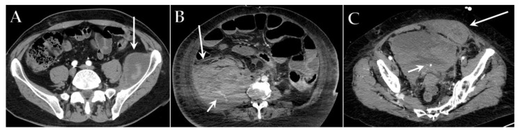
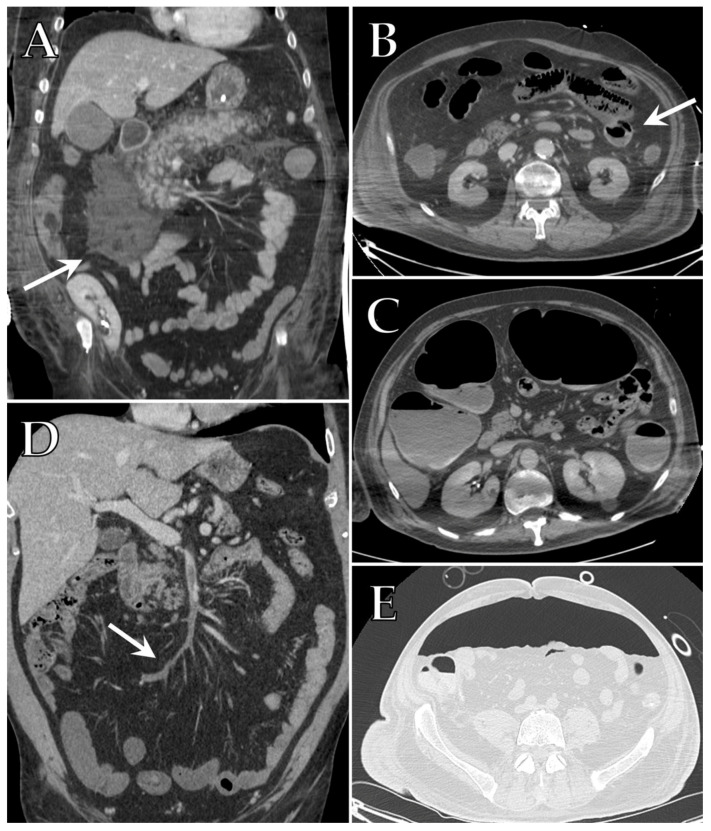
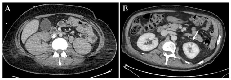
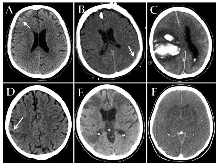
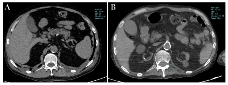
A significant proportion of patients with COVID-19 pneumonia could develop acute respiratory distress syndrome (ARDS), thus requiring mechanical ventilation, and resulting in a high rate of intensive care unit (ICU) admission. Several complications can arise during an ICU stay, from both COVID-19 infection and the respiratory supporting system, including barotraumas (pneumothorax and pneumomediastinum), superimposed pneumonia, coagulation disorders (pulmonary embolism, venous thromboembolism, hemorrhages and acute ischemic stroke), abdominal involvement (acute mesenteric ischemia, pancreatitis and acute kidney injury) and sarcopenia. Imaging plays a pivotal role in the detection and monitoring of ICU complications and is expanding even to prognosis prediction. The present pictorial review describes the clinicopathological and radiological findings of COVID-19 ARDS in ICU patients and discusses the imaging features of complications related to invasive ventilation support, as well as those of COVID-19 itself in this particularly fragile population. Radiologists need to be familiar with COVID-19's possible extra-pulmonary complications and, through reliable and constant monitoring, guide therapeutic decisions. Moreover, as more research is pursued and the pathophysiology of COVID-19 is increasingly understood, the role of imaging must evolve accordingly, expanding from the diagnosis and subsequent management of patients to prognosis prediction.

Keywords: ARDS; COVID-19; intensive care; lung CT; mechanical intubation; pneumomediastinum; pneumothorax; pulmonary embolism; superinfection.


Conflict of interest statement

The authors declare no conflict of interest.

Figures

Figure 1
Figure 1
Axial HRCT images of a 38-year-old man with COVID-19 ARDS admitted to ICU at the same level, performed at different times: baseline scan (A) and 7-month follow-up (B). The baseline scan (A) shows typical imaging features indicative of severe COVID-19 pneumonia, including extensive bilateral parenchymal consolidations, mainly affecting the posterior regions of lower lobes, bilateral focal ground-glass opacities in the anterior regions and patchy consolidation, peripherally distributed, resembling pulmonary fibrosis. The 7-month scan (B) shows a complete resolution of the parenchymal consolidations and the apparent fibrotic abnormalities.
Figure 2
Figure 2
Axial HRCT images of different patients with COVID-19 ARDS admitted to ICU showing imaging features indicative of barotrauma secondary to mechanical ventilation: (A) large pneumatocele (black arrow); (B) small anterior pneumothorax (black arrow); (C) large left pneumothorax; (D) extensive bilateral subcutaneous emphysema of the neck; (E) pneumomediastinum; (F) pneumomediastinum associated with small anterior pneumothorax (small black arrow) and subcutaneous emphysema of the left upper chest (long black arrow).
Figure 3
Figure 3
Axial HRCT images of different patients admitted to ICU demonstrating a sudden increase of large consolidations (black arrows) (A,B) associated with worsening of COVID-19 pneumonia. Superimposed infections of Acinetobacter (A) and Aspergillus (B) were detected. A different COVID-19 patient showed pulmonary consolidation complicated by cavitation located in the medium lobe (black arrow) (C), which demonstrates communication with the bronchial tree at minimum intensity projection (MIP) reconstruction (black arrow) (D); superinfection of Pseudomonas aeruginosa was detected.
Figure 4
Figure 4
Axial contrast-enhanced CT images of different ICU patients with COVID-19 show filling defects indicative of a massive lung embolism in the right pulmonary artery (white arrow) (A), multiple embolisms in the left lower lobe artery (white arrow) (B), thrombosis of the left jugular vein (white arrow) (C) and thrombosis of the superior vena cava near the central venous catheter (white arrow) (D).
Figure 5
Figure 5
Axial contrast-enhanced CT images of different patients with COVID-19 admitted to ICU that developed hemorrhagic events: (A) a hematoma of the left iliopsoas muscle (white arrow); (B) a large retroperitoneal hematoma (long white arrow) that displaces the small bowel and shows a blush of active arterial bleeding (small white arrow); (C) a spontaneous hematoma of the anterior rectus abdominus (long white arrow) collecting in the pelvis that shows a small blush of active arterial bleeding (small white arrow).
Figure 6
Figure 6
Coronal contrast-enhanced CT image of a patient with COVID-19 admitted to ICU with suspected pancreatitis reveals a diffusely enlarged pancreas, with ill-defined borders, stranding of the peri-pancreatic fat and streak of fluid along the anterior conal fascia (white arrow) (A). Axial contrast-enhanced CT scan of different COVID-19 patients with suspected acute mesenteric ischemia demonstrating the presence of linear collections of gas inside the small bowel wall referable to pneumatosis intestinalis (white arrow) (B) and dilated intestinal loops with a paper-thin wall and air-fluid levels (C). Coronal contrast-enhanced CT image of a COVID-19 patient admitted to ICU showing thrombosis of the superior mesenteric vein extending to several jejuno-ileal branches (white arrow), with consequent loss of contrast enhancement of the corresponding vascularized loops (D). Axial unenhanced CT scan with lung window of an ICU patient with COVID-19 and abdominal pain, showing a large quantity of free air (E).
Figure 7
Figure 7
Axial contrast-enhanced CT image of an ICU patient with COVID-19 ARDS in ECMO treatment that developed kidney injury, showing enlarged kidney with a loss of corticomedullary differentiation (A). Axial contrast-enhanced CT image of a different patient with severe COVID-19 admitted to ICU demonstrating a wedge-shaped region of hypoattenuation in the left kidney (white arrow) consistent with renal infarction (B).
Figure 8
Figure 8
Axial unenhanced CT images of different patients with severe COVID-19 admitted to ICU showing spontaneous subacute right frontoparietal subdural hematoma (white arrow) (A), acute left parieto-occipital subdural hematoma (white arrow) (B) and massive right intracerebral hemorrhage with surrounding edema and significant midline shift (C). Axial unenhanced CT images show an acute ischemic stroke in the right parietal lobe (white arrow) (D) and multiple bilateral acute ischemic strokes (E) in two different COVID-19 patients admitted to ICU. Axial contrast-enhanced CT of a COVID-19 comatose patient reveals diffuse sulcal effacement with a decreased differentiation between gray and white matter, compatible with severe anoxic brain injury (F).
Figure 9
Figure 9
Axial unenhanced CT images of a patient with severe COVID-19 at the same level (T12), performed at the time of the ICU admission (A) and after a 3-month of ICU stay (B), demonstrates a reduction in muscle mass in the left paravertebral muscle (i.e., <30 HU) during hospitalization.

References

    1. Simpson S., Kay F.U., Abbara S., Bhalla S., Chung J.H., Chung M., Henry T.S., Kanne J.P., Kligerman S., Ko J.P., et al. Radiological Society of North America Expert Consensus Statement on Reporting Chest CT Findings Related to COVID-19. Endorsed by the Society of Thoracic Radiology, the American College of Radiology, and RSNA—Secondary Publication. J. Thorac. Imaging. 2020;35:219–227. doi: 10.1097/RTI.0000000000000524. - DOI - PMC - PubMed
    1. Balacchi C., Brandi N., Ciccarese F., Coppola F., Lucidi V., Bartalena L., Parmeggiani A., Paccapelo A., Golfieri R. Comparing the first and the second waves of COVID-19 in Italy: Differences in epidemiological features and CT findings using a semi-quantitative score. Emerg. Radiol. 2021;28:1055–1067. doi: 10.1007/s10140-021-01937-y. - DOI - PMC - PubMed
    1. Ciccarese F., Coppola F., Spinelli D., Galletta G.L., Lucidi V., Paccapelo A., DE Benedittis C., Balacchi C., Golfieri R. Diagnostic Accuracy of North America Expert Consensus Statement on Reporting CT Findings in Patients Suspected of Having COVID-19 Infection: An Italian Single-Center Experience. Radiol. Cardiothorac. Imaging. 2020;2:e200312. doi: 10.1148/ryct.2020200312. - DOI - PMC - PubMed
    1. Angeli E., Dalto S., Marchese S., Setti L., Bonacina M., Galli F., Rulli E., Torri V., Monti C., Meroni R., et al. Prognostic value of CT integrated with clinical and laboratory data during the first peak of the COVID-19 pandemic in Northern Italy: A nomogram to predict unfavorable outcome. Eur. J. Radiol. 2021;137:109612. doi: 10.1016/j.ejrad.2021.109612. - DOI - PMC - PubMed
    1. Tabatabaei S.M.H., Talari H., Moghaddas F., Rajebi H. CT Features and Short-term Prognosis of COVID-19 Pneumonia: A Single-Center Study from Kashan, Iran. Radiol. Cardiothorac. Imaging. 2020;2:e200130. doi: 10.1148/ryct.2020200130. - DOI - PMC - PubMed

LinkOut - more resources