. 2022 Mar 31;12(4):873.
doi: 10.3390/diagnostics12040873.

Prenatal Diagnosis and Outcome of Umbilical-Portal-Systemic Venous Shunts: Experience of a Tertiary Center and Proposal for a New Complex Type

Affiliations

Prenatal Diagnosis and Outcome of Umbilical-Portal-Systemic Venous Shunts: Experience of a Tertiary Center and Proposal for a New Complex Type

Rodica Daniela Nagy et al. Diagnostics (Basel). .

Abstract

Aims: To share our experience in the prenatal diagnosis of umbilical-portal-systemic venous shunts (UPSVS) and to study the prognostic factors for proper prenatal and perinatal management. Material and Methods: A five-year prospective study regarding the detection of UPSVS was conducted in two referral centers, Medgin Ginecho Clinic and the Prenatal Diagnostic Unit of the tertiary center, University Emergency County Hospital Craiova, Romania. We included in the analysis a series of agenesis of ductus venosus (ADV) cases previously reported by our center. We analyzed the incidence of the UPSVS types, their associations, and outcome predictors. Results: UPSVS were diagnosed in all 16 cases that were presented to our center at the time of first trimester anomaly scan, except one (94.12%). We diagnosed: 19 type I (61.2%), 4 type II (12.9%) and 5 type IIIa (16.1%) UPSVS. In three cases (9.6%) we noted multiple shunts, which we referred to as type IV (a new UPSVS type). Type IIIa-associated fetal growth restriction (FGR) was found in 60% of cases. Major anomalies worsened the outcome. Of the UPVSS cases, 57.1% were associated with PVS anomalies. Genetic anomalies were present in 40% of the tested cases. Conclusions: The incidence of UPSVS in our study was 0.2%. Early detection is feasible. The postnatal outcome mainly depends on the presence of structural, genetic and PVS anomalies. FGR may be associated. The new category presented a poor outcome secondary to poor hemodynamic and major associated anomalies.

Keywords: agenesis of ductus venosus; color Doppler; fetal venous shunt; portal system; prenatal diagnosis; umbilical drainage; umbilical–portal–systemic venous shunt; venous anomalies.


Conflict of interest statement

The authors declare no conflict of interest.

Figures

Figure 1
Figure 1
The typical aspect of the portal venous system (PVS), hepatic veins, and ductus venosus. (A) Transverse plane of the fetal abdomen, with high-definition directional power Doppler, applied to demonstrate the normal L-shaped UV confluence and PVS features. (B) Transverse plane allowed the evaluation of the left middle and right hepatic veins, aorta, and inferior vena cava. (C) Sagittal plane of the fetal thorax and abdomen, with the evaluation of the umbilical vein, left hepatic vein, ductus venosus, and inferior vena cava. UV, umbilical vein; RAPV, anterior branch of the right portal vein; RPPV, posterior branch of the right portal vein; LiPV, left portal vein inferior branch; LmPV, left portal vein medial branch; Ao, aorta; RHV, right hepatic vein; LHV, left hepatic vein; MHV, middle hepatic vein.
Figure 2
Figure 2
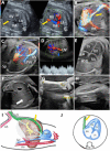
UPSVS type I with IVC drainage of the umbilical vein at 13 weeks of gestation (Case 1). (A) Color Doppler imaging in the longitudinal plane, showing the umbilical vein draining into the inferior vena cava. (B) Four-dimensional STIC reconstruction presenting the abnormal drainage of the umbilical vein (red arrow). (C) Four-dimensional STIC reconstruction, top view, showing the medial and superior branches of the left portal vein and the inferior vena cava drainage. (D) Axial view at the level of the fetal abdomen demonstrating the absence of the right portal vein and the umbilical vein connection to the inferior vena cava. Blue mark—skin edema, yellow mark—ascites. (E) Two-dimensional axial view at the thorax level showing the right-sided heart due to the presence of the pleural effusion (red mark). (F) Four-chamber view. During diastole, atrioventricular flows are visualized, separated by the interatrial and interventricular septum. (G) Color Doppler imaging, showing the pulmonary artery crossing the aorta in the axial plane. (H) Three vessels and trachea view showing the superior vena cava, pulmonary artery, ductus arteriosus, transverse aortic arch, and trachea. SVC, superior vena cava; Ao, aorta; PA, pulmonary artery; DA, ductus arteriosus; Dao, descending aorta; Tr, trachea. IVC, inferior vena cava; Sp, spine; RA, right atrium; LA, left atrium; RV, right ventricle; LV, left ventricle; P, lungs; UV, umbilical vein; HV, hepatic vein; Ao, aorta; LPVs, the superior branch of the left portal vein; LPVm, medial branch of the left portal vein; St, stomach.
Figure 3
Figure 3
UPSVS type I with cardiac drainage of the umbilical vein at 24 weeks of gestation (Case 3). (A) Umbilical drainage (yellow arrow) directly to the right atrium visible in an axial–oblique plane of the upper abdomen and low thorax region, using duplex (color Doppler and gray-scale) imaging. (B) Directional power Doppler applied in the sagittal plane of the upper abdomen and low thorax region, showing the umbilical vein abnormal connection to the right atrium and absence of the ductus venosus. (C) Oblique plane at the abdomen level showing the absence of right portal vein. (D) Typical triphasic aspect of the shunt. (E) Four-chamber view, which demonstrates the absence of septum secundum. (F) The axial abdominal sweep could not find the gallbladder (white arrow). (G) Two-dimensional evaluation showing unilateral absence of the radius. (H) Short humerus. (I,J) Schematic diagram of type I shunt. (I) Sagittal plane with normal course of umbilical–portal–DV complex and hepatic veins observed by the transparency of the oval and also the abnormal course of the umbilical vein draining into the right atrium. (J) Four-chamber view, which demonstrates the abnormal drainage of the umbilical vein. IVC, inferior vena cava; Sp, spine; RA, right atrium; LA, left atrium; RV, right ventricle; LV, left ventricle; LPVs, the superior branch of the left portal vein; LPVi, the inferior branch of the left portal vein; ASD, atrial septal defect; UV, umbilical vein; Ao, aorta; HV, hepatic vein; PS portal sinus; RPV, right portal vein; CT, celiac trunk; DV, ductus venosus; UA, umbilical artery.
Figure 4
Figure 4
UPSVS type II with hepatic vein drainage of the umbilical vein at 13 weeks of gestation (Case 11). (A,B) In the sagittal plane, color Doppler imaging shows the umbilical vein draining through a short ductus venosus-like vessel (arrow) into the hepatic. (C) Four-dimensional STIC reconstruction, showing in the sagittal plane the abnormal drainage of the umbilical vein (blue circle). (D) Four-chamber view. During diastole, flow is visualized entering from the right and left atria (RA, LA) into the right and left ventricles (RV, LV), and the interatrial and interventricular septum separates the flows. (E) Normal “V-sign” with pulmonary artery, ductus arteriosus, and transverse aortic arch. (F) Midsagittal plane of the fetal head and upper thorax, showing normal appearance of the first-trimester genetic markers: nuchal translucency (NT) and nasal bone. (G,H) Schematic diagram of type II shunt. (G) Sagittal and (H) axial plane with abnormal course of umbilical vein draining into the hepatic vein through a ductus venosus-like vessel. SVC, superior vena cava; Ao, aorta; MPA, main pulmonary artery; DA, ductus arteriosus; Dao, descending aorta; Tr, trachea. IVC, inferior vena cava; Sp, spine; RA, right atrium; LA, left atrium; RV, right ventricle; LV, left ventricle; UV, umbilical vein; HV, hepatic vein; MHV, medial branch of the hepatic vein; RHV, right hepatic vein; LHV, left hepatic vein; Ao, aorta; RPV, right portal vein; LPV, left portal vein; PS portal sinus; RPV, right portal vein; DV, ductus venosus; UA, umbilical artery.
Figure 5
Figure 5
Type IIIa shunt with intrahepatic drainage of the umbilical vein at 30 weeks of gestation (Case 16) (A) Color Doppler imaging, showing in the oblique–transverse plane the connection between the superior branch of the left portal vein and the left hepatic vein (B) Four-dimensional STIC rendering of the abdominal vessels showing the pathway of the shunt. (C) Transverse view at the level of insertion of the umbilical cord showing intrahepatic drainage of the shunt and the biphasic aspect of the velocity. (D) Four-chamber view color Doppler assessment with flows separated by the interatrial and interventricular septum. (E) Color Doppler imaging, showing the left ventricular outflow tract in the axial plane. (F) Three vessels and trachea view showing the superior vena cava, pulmonary artery, ductus arteriosus, transverse aortic arch (from the proximal aorta to the descending aorta), and trachea. (G) Transverse plane of the fetal abdomen showing the typical UV confluence and portal venous system features. (H) Two-dimensional imaging of the hyper-coiled umbilical cord. (I) Graph of the fetal weight demonstrating a low percentile for the gestational age. (J,K) Schematic diagram of type III shunt. (J) Sagittal and (K) axial plane with abnormal connection between the superior branch of the left portal vein and the left hepatic vein. LPVi, the inferior branch of the left portal vein; LPVm, medial branch of the left portal vein; IVC, inferior vena cava; UV, umbilical vein; HVr, right branch of the hepatic vein; HVm, medial branch of the hepatic vein; HVl, left branch of the hepatic vein; Ao, aorta; Sp, spine.
Figure 6
Figure 6
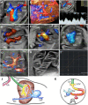
Complex drainage of the umbilical vein (UV) into the hepatic vein (HV) and two other intrahepatic shunts (one between the shunt and the left hepatic vein and the other between the right portal vein and the IVC) at 18 and 28 weeks of gestation (WG) (Case 18). (A) Color Doppler assessment at 18 WG showing the drainage of the UV into the middle branch of the HV. (B) Four-dimensional STIC rendering showing in the sagittal plane the short shunt vessel (yellow circle) connecting the umbilical and hepatic vein, below the prediaphragmatic infundibulum. (C) Four-dimensional STIC rendering showing two abnormal connections: one between the UV and the HV (white arrows) and another between the right portal vein and the IVC (yellow arrows). (DG) Consecutive axial abdominal planes, part of the umbilical–portal and hepatic circulation investigation. (D) Caudally, a transverse plane at the level of the portal sinus. (E) A connection between the right portal vein and IVC (yellow arrow) is identified in the upper abdomen. (F) The white circle highlights a connection between the left hepatic vein and an abnormal shunt between one smaller venous branch of the portal vein (red arrow) and the umbilical vein. (G) Axial plane at the level of the hepatic efferent venous system and the umbilical–hepatic shunt (yellow circle). (HK) The four-chamber and outflow tract views along with massive pleural effusion (yellow mark). (L) Ultrasound image of septated cystic hygroma at 18 WG. (M) Transverse plane at the level of the fetal abdomen with the typical aspect of the portal venous system. (N) Lateral view of the heart with hypertrophic ventricular walls and interventricular septum (blue arrows) and the presence of pleural effusion (white arrows). (O) Axial oblique posterior fossa view showing small cerebellar hemispheres. (P,Q) Schematic diagram of type IV shunt. (P) Sagittal and (Q) axial plane with abnormal course of umbilical vein draining into the hepatic vein and abnormal connection between right portal vein and inferior vena cava. RAPV, anterior branch of the right portal vein; RPPV, the posterior branch of the right portal vein; MPV, main portal vein; LPVs, the superior branch of the left portal vein. LPVi, the inferior branch of the left portal vein; LPVm, medial branch of the left portal vein; IVC, inferior vena cava; UV, umbilical vein; HV, hepatic vein; LHV, left branch of the hepatic vein; MHV, medial branch of the hepatic vein; RHV, right branch of the hepatic vein; LA, left atrium; LV, left ventricle; RA, right atrium; RV, right ventricle; St, stomach; Ao, aorta.

Similar articles

Cited by

References

    1. Fasouliotis S.J., Achiron R., Kivilevitch Z., Yagel S. The human fetal venous system: Normal embryologic, anatomic, and physiologic characteristics and developmental abnormalities. J. Ultrasound Med. Off. J. Am. Inst. Ultrasound Med. 2002;21:1145–1158. doi: 10.7863/jum.2002.21.10.1145. - DOI - PubMed
    1. Stringer M.D. The clinical anatomy of congenital portosystemic venous shunts. Clin.Anat. Off. J. Am. Assoc. Clin. Anat. Br. Assoc. Clin. Anatomists. 2008;21:147–157. doi: 10.1002/ca.20574. - DOI - PubMed
    1. Grazioli L., Alberti D., Olivetti L., Rigamonti W., Codazzi F., Matricardi L., Fugazzola C., Chiesa A. Congenital absence of portal vein with nodular regenerative hyperplasia of the liver. Eur. Radiol. 2000;10:820–825. doi: 10.1007/s003300051012. - DOI - PubMed
    1. Kanamori Y., Hashizume K., Kitano Y., Sugiyama M., Motoi T., Tange T. Congenital extrahepatic portocaval shunt (Abernethy type 2), huge liver mass, and patent ductus arteriosus—A case report of its rare clinical presentation in a young girl. J. Pediatr. Surg. 2003;38:E15. doi: 10.1053/jpsu.2003.50153. - DOI - PubMed
    1. Murray C.P., Yoo S.J., Babyn P.S. Congenital extrahepatic portosystemic shunts. Pediatr. Radiol. 2003;33:614–620. - PubMed