

Comparison of RetCam and Smartphone-Based Photography for Retinopathy of Prematurity Screening
- PMID: 35453993
- PMCID: PMC9029155
- DOI: 10.3390/diagnostics12040945
Comparison of RetCam and Smartphone-Based Photography for Retinopathy of Prematurity Screening
Abstract
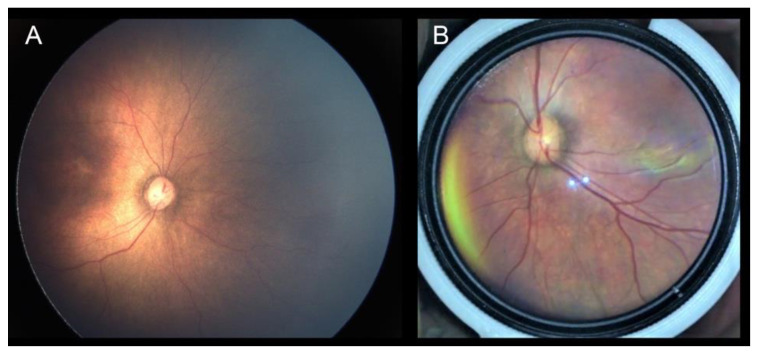
This study aimed to compare the clinical performance between a smartphone-based fundus photography device and a contact imaging device for retinopathy of prematurity (ROP) screening. All patients were first examined with binocular indirect ophthalmoscopy (BIO), which served as the reference standard. The patients were then assessed by two devices. Imaging quality, ability to judge the zone and stage of ROP, agreement with the BIO results, vital signs, and pain scores were compared between these two devices. In total, 142 eyes of 71 infants were included. For the smartphone-based fundus photography, image quality was graded excellent or acceptable in 91.4% of examinations, although it was still significantly inferior to that of the contact imaging device (p < 0.001). The smartphone-based fundus photography images had moderate agreement with the BIO results regarding the presence or absence of plus disease (Cohen’s κ = 0.619), but evaluating the zone (p < 0.001) and stage (p < 0.001) of ROP was difficult. Systemic parameters, except for heart rate, were similar between the two imaging devices (all p > 0.05). In conclusion, although the smartphone-based fundus photography showed moderate agreement for determining the presence or absence of plus disease, it failed to identify the zone and stage of ROP.
Keywords: imaging systems; retinopathy of prematurity; smartphone-based screen; telescreening.
Conflict of interest statement
The authors declare no conflict of interest.
Figures


References
-
- Palmer E.A., Hardy R.J., Dobson V., Phelps D.L., Quinn G.E., Summers C.G., Krom C.P., Tung B. 15-year outcomes following threshold retinopathy of prematurity: Final results from the multicenter trial of cryotherapy for retinopathy of prematurity. Arch. Ophthalmol. 2005;123:311–318. doi: 10.1001/archopht.123.3.311. - DOI - PubMed
Grants and funding
LinkOut - more resources
Full Text Sources
