Review
. 2022 Apr 12;12(4):955.
doi: 10.3390/diagnostics12040955.

Parenchymal Insults in Abuse-A Potential Key to Diagnosis

Affiliations
Review

Parenchymal Insults in Abuse-A Potential Key to Diagnosis

Marguerite M Caré. Diagnostics (Basel). .

Abstract

Subdural hemorrhage is a key imaging finding in cases of abusive head trauma and one that many radiologists and radiology trainees become familiar with during their years of training. Although it may prove to be a marker of trauma in a young child or infant that presents without a history of injury, the parenchymal insults in these young patients more often lead to the debilitating and sometimes devastating outcomes observed in this young population. It is important to recognize these patterns of parenchymal injuries and how they may differ from the imaging findings in other cases of traumatic injury in young children. In addition, these parenchymal insults may serve as another significant, distinguishing feature when making the medical diagnosis of abusive head injury while still considering alternative diagnoses, including accidental injury. Therefore, as radiologists, we must strive to look beyond the potential cranial injury or subdural hemorrhage for the sometimes more subtle but significant parenchymal insults in abuse.

Keywords: abusive head trauma; child abuse; computed tomography; hypoxic-ischemic injury; magnetic resonance imaging.


Conflict of interest statement

The author declares no conflict of interest.

Figures
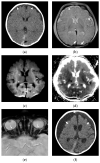
Figure 1
Figure 1
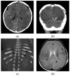
Subdural hemorrhage and subdural collections in an unresponsive 2-month-old male presenting with forehead bruising and respiratory depression. (a) Axial noncontrast head CT image demonstrates thin, high attenuation subdural hemorrhage along the posterior left occipital lobe (black arrow) and bilateral, low attenuation frontal subdural collections (white arrowheads). There is also subtle loss of gray-white matter differentiation bilaterally; (b) axial T2-weighted MR image on day 4 shows regions of decreased gray-white matter differentiation throughout both cerebral hemispheres with T2 hyperintense subdural collections (white arrowheads) and hypointense subdural hemorrhage posteriorly (white arrows); (c) axial b-1000 diffusion-weighted image demonstrates diffuse areas of abnormal, restricted diffusion (white arrowheads) in both cerebral hemispheres consistent with cytotoxic edema/hypoxic-ischemic injury; (d) apparent diffusion coefficient image shows corresponding regions of diffusion restriction predominantly in cortical and subcortical regions of both cerebral hemispheres (black arrowheads); (e) axial susceptibility-weighted image shows small, bilateral retinal hemorrhages (white arrows), confirmed clinically; (f) axial CT image at 2 month follow-up show diffuse brain parenchymal volume loss with now large, bilateral subdural collections (white arrows).
Figure 2
Figure 2
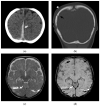
Diffuse, bilateral hemispheric attenuation abnormality in a 3-month-old male presenting in status epilepticus with multifocal facial, extremity, and trunk bruises. (a) Initial axial noncontrast head CT image with diffuse, abnormal attenuation throughout both cerebral hemispheres (white arrowheads); (b) coronal noncontrast CT image shows thin subdural hemorrhage along the left tentorial leaflet (white arrow); (c) coronal reconstruction bone algorithm image from an abdominal CT at presentation shows healing, bilateral posterior rib fractures (black arrowheads); (d) axial T2-weighted image on day 4 demonstrates diffuse, hemispheric loss of gray-white matter differentiation (black arrowheads); (e) axial b-1000 diffusion-weighted image demonstrates diffuse areas of abnormal, restricted diffusion (black arrows) in both cerebral hemispheres consistent with cytotoxic edema/hypoxic-ischemic injury; (f) apparent diffusion coefficient image shows corresponding regions of diffusion restriction predominantly in cortical and subcortical regions of both cerebral hemispheres (black arrows).
Figure 2
Figure 2
Diffuse, bilateral hemispheric attenuation abnormality in a 3-month-old male presenting in status epilepticus with multifocal facial, extremity, and trunk bruises. (a) Initial axial noncontrast head CT image with diffuse, abnormal attenuation throughout both cerebral hemispheres (white arrowheads); (b) coronal noncontrast CT image shows thin subdural hemorrhage along the left tentorial leaflet (white arrow); (c) coronal reconstruction bone algorithm image from an abdominal CT at presentation shows healing, bilateral posterior rib fractures (black arrowheads); (d) axial T2-weighted image on day 4 demonstrates diffuse, hemispheric loss of gray-white matter differentiation (black arrowheads); (e) axial b-1000 diffusion-weighted image demonstrates diffuse areas of abnormal, restricted diffusion (black arrows) in both cerebral hemispheres consistent with cytotoxic edema/hypoxic-ischemic injury; (f) apparent diffusion coefficient image shows corresponding regions of diffusion restriction predominantly in cortical and subcortical regions of both cerebral hemispheres (black arrows).
Figure 3
Figure 3
Mixed attenuation right subdural hemorrhage in a 3-year-old female fatality presenting with abrupt mental status change and multifocal bruises after a reported fall. (a) Initial axial noncontrast head CT demonstrates a mixed attenuation subdural hemorrhage (white arrow) with mass effect, midline shift (black arrowhead), and effaced right temporal horn (black arrow); (b) next-day CT image shows changes of a decompressive hemicraniectomy with diffuse, abnormal attenuation of the right cerebral hemisphere (white arrow), which herniates through the cranial defect.
Figure 4
Figure 4
Axonal injury and lacerations/contusional tears in a 4-month-old male presenting with seizure and lethargy. (a) Initial axial CT image demonstrates right parafalcine high-attenuation subdural hemorrhage (white arrow); (b) coronal reconstruction in bone algorithm demonstrates a right parietal bone fracture (black arrow) with overlying soft tissue swelling (white arrowhead); (c) axial T2-weighted MR image on day 4 demonstrates bilateral lacerations/contusional tears with fluid-hemorrhagic levels near the temporal-occipital lobe junctions (white arrows); (d) axial susceptibility-weighted image demonstrates layering hemorrhage in lacerations/contusional tears (white arrows) and axonal injury near the cortical-white matter junction in each frontal lobe (black arrows).
Figure 5
Figure 5
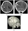
Skull fracture and hemorrhagic contusion in a 2-month-old female presenting with the history of a fall off a couch. (a) Initial axial CT demonstrates a hemorrhagic contusion at the posterior temporal-parietal lobe junction (white arrow) with mixed but predominantly low-attenuation hemorrhagic subdural (black arrow). More focal loss of gray-white matter differentiation (black arrowhead) may also suggest coexistent venous infarction or bland contusion; (b) coronal CT reconstruction shows the hemorrhagic contusion (white arrow) deep to a diastatic right parietal bone fracture (white arrowhead); (c) three-dimensional reconstruction of the skull demonstrates a mildly complex right parietal bones fracture (black arrows) and right frontal bone fracture (white arrow).
Figure 6
Figure 6
Focal laceration or contusional tear in a 2-month-old former preterm male infant presenting with seizure. (a) Initial axial noncontrast CT demonstrates a small, focal, left frontal lobe laceration/tear with layering posterior hemorrhage (white arrow); (b) axial T1-weighted image shows a hypointense, well defined laceration/tear with subtle layering hemorrhage posteriorly (white arrow); (c) axial multiplanar gradient recalled acquisition in the steady-state (MPGR) image shows the layering posterior hemorrhage (white arrow); (d) follow-up skeletal survey demonstrates healing right acromion fracture (black arrow) and distal humeral periosteal reaction (white arrow) suggesting a healing fracture.
Figure 6
Figure 6
Focal laceration or contusional tear in a 2-month-old former preterm male infant presenting with seizure. (a) Initial axial noncontrast CT demonstrates a small, focal, left frontal lobe laceration/tear with layering posterior hemorrhage (white arrow); (b) axial T1-weighted image shows a hypointense, well defined laceration/tear with subtle layering hemorrhage posteriorly (white arrow); (c) axial multiplanar gradient recalled acquisition in the steady-state (MPGR) image shows the layering posterior hemorrhage (white arrow); (d) follow-up skeletal survey demonstrates healing right acromion fracture (black arrow) and distal humeral periosteal reaction (white arrow) suggesting a healing fracture.

Similar articles

Cited by

References

    1. U.S. Department of Health and Human Services, AfCaF Child Welfare Information Gateway. Child Abuse and Neglect Fatalities 2019: Statistics and Interventions. [(accessed on 28 February 2022)];2021 Available online: https://www.childwelfare.gov/pubs/factsheets/fatality/
    1. Palusci V.J., Covington T.M. Child maltreatment deaths in the U.S. National Child Death Review Case Reporting System. Child Abus. Negl. 2014;38:25–36. doi: 10.1016/j.chiabu.2013.08.014. - DOI - PubMed
    1. Jackson J.E., Beres A.L., Theodorou C.M., Ugiliweneza B., Boakye M., Nuño M. Long-term impact of abusive head trauma in young children: Outcomes at 5 and 11 years old. J. Pediatr. Surg. 2021;56:2318–2325. doi: 10.1016/j.jpedsurg.2021.02.019. - DOI - PMC - PubMed
    1. Chevignard M.P., Lind K. Long-term outcome of abusive head trauma. Pediatr. Radiol. 2014;44((Suppl. S4)):S548–S558. doi: 10.1007/s00247-014-3169-8. - DOI - PubMed
    1. Choudhary A.K., Servaes S., Slovis T.L., Palusci V.J., Hedlund G.L., Narang S.K., Moreno J.A., Dias M.S., Christian C.W., Nelson M.D., Jr., et al. Consensus statement on abusive head trauma in infants and young children. Pediatr. Radiol. 2018;48:1048–1065. doi: 10.1007/s00247-018-4149-1. - DOI - PubMed

LinkOut - more resources