Review
. 2022 Apr 12;12(4):969.
doi: 10.3390/diagnostics12040969.

Nucleic Acid Biomarkers in Waldenström Macroglobulinemia and IgM-MGUS: Current Insights and Clinical Relevance

Affiliations
Review

Nucleic Acid Biomarkers in Waldenström Macroglobulinemia and IgM-MGUS: Current Insights and Clinical Relevance

Daniela Drandi et al. Diagnostics (Basel). .

Abstract

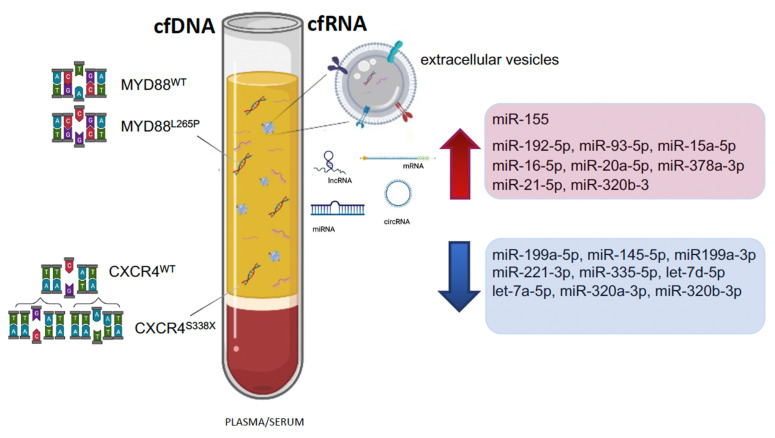
Waldenström Macroglobulinemia (WM) is an indolent lymphoplasmacytic lymphoma, characterized by the production of excess immunoglobulin M monoclonal protein. WM belongs to the spectrum of IgM gammopathies, ranging from asymptomatic IgM monoclonal gammopathy of undetermined significance (IgM-MGUS), through IgM-related disorders and asymptomatic WM to symptomatic WM. In recent years, its complex genomic and transcriptomic landscape has been extensively explored, hereby elucidating the biological mechanisms underlying disease onset, progression and therapy response. An increasing number of mutations, cytogenetic abnormalities, and molecular signatures have been described that have diagnostic, phenotype defining or prognostic implications. Moreover, cell-free nucleic acid biomarkers are increasingly being investigated, benefiting the patient in a minimally invasive way. This review aims to provide an extensive overview of molecular biomarkers in WM and IgM-MGUS, considering current shortcomings, as well as potential future applications in a precision medicine approach.

Keywords: CXCR4; IgM-MGUS; MYD88; WM; cfDNA; liquid biopsy; lncRNA; miRNA.


Conflict of interest statement

The authors declare no conflict of interest.

Figures

Figure 1
Figure 1
Diagnostic criteria for WM and IgM-MGUS. Serum monoclonal IgM: any concentration according to Owen 2003 (2° IWWM), Swerdlow 2008 (WHO 2008) and Campo 2011 (WHO 2011) [9,11,13] or ≥30 g/L according to Ansell 2010 (mSMART), Swerdlow 2017 and Maqbool 2020 [14,17,19]. BM infiltration: unequivocal BM infiltration by lymphoplasmacytic lymphoma [9,11] or infiltration ≥10% [14,17,19]. Immunophenotype: immunophenotype consistent with WM: CD19+, CD20+, CD22+, CD79+, CD5-, CD10-, CD23-. Of note, 5–10% of WM cases could express CD5 [20,21]. Symptoms: attributable to tumor infiltration (in BM or extramedullary) and/or to the monoclonal protein (related to immunological or chemical properties) [7,8,21]. IgM-related disorders: patients who have clinical features attributable to the IgM monoclonal protein but without overt evidence of LPL in the BM. For cases in which BM infiltration is not confirmed, the immunophenotypic profile is useful to discriminate the pattern of WM from other IgM-related disorders. However, BM infiltration by immunohistochemistry is mandatory for a definitive WM diagnosis. IgM-MGUS: patients with an IgM gammopathy, without evidence of LPL in the BM biopsy and no symptoms. Cases with detectable clonal B cells by immunophenotype and absence of BM infiltration by LPL [9] or BM infiltration <10% and IgM <30 g/L [14,17,19] should be classified as IgM-MGUS. aWM: patients with an IgM gammopathy and BM infiltration by LPL without symptoms. Immunophenotyping is strongly recommended for differential diagnosis. WM: patients with IgM protein of any concentration and unequivocal BM infiltration and symptoms. Immunophenotyping is strongly recommended.
Figure 2
Figure 2
Mutational and cytogenetic landscape of WM. The figure describes the association between genomic abnormalities in WM patients. (A) From the center outward, the distribution and overlap of MYD88L265P mutations, CXCR4MUT mutations, karyotype (K) (Complex K: <5 clonal aberrations; high complex K: ≥5 clonal aberrations), copy number alterations (CNAs) and less frequent mutations (MUTs) are shown, respectively. The color code in the outer ring refers to colors in panels B and C. Relevant up and down-regulated genes (arrows) are reported. DLBCL like mutations: somatic mutations overlapping those detected in diffuse large B cell lymphoma (DLBCL), (i.e., TBL1XR1, PTPN13, MALT1, BCL10, NFKB2, NFKBIB, NFKBIZ, and UDRL1F). (B) Relative distribution of cytogenetic abnormalities. tri: trisomy, del: deletion. (C) Relative distribution of uncommon mutations. The percentage (%) of the most frequent MUTs and CNAs are estimated based on published data, for more details see below.
Figure 3
Figure 3
An illustrative overview of the pathways involved with increased miR-155 expression in WM. MAFB, SHANK2 and SH3PXD2A (italic) are more recently discovered targets of miR-155 in WM and further studies are needed to elucidate their role. Everolimus-dependent anti-WM activity is partially driven by targeting miR-155 (red).
Figure 4
Figure 4
Cell-free DNA (cfDNA) and cell-free RNA (cfRNA) markers in plasma or serum of WM patients. Significant up- and downregulated miRNAs are shown (arrows).

Similar articles

Cited by

References

    1. Ziegler A., Koch A., Krockenberger K., Grosshennig A. Personalized medicine using DNA biomarkers: A review. Hum. Genet. 2012;131:1627–1638. doi: 10.1007/s00439-012-1188-9. - DOI - PMC - PubMed
    1. Gilboa T., Garden P.M., Cohen L. Single-molecule analysis of nucleic acid biomarkers—A review. Anal. Chim. Acta. 2020;1115:61–85. doi: 10.1016/j.aca.2020.03.001. - DOI - PubMed
    1. Braggio E., Philipsborn C., Novak A., Hodge L., Ansell S., Fonseca R. Molecular pathogenesis of Waldenstrom’s macroglobulinemia. Haematologica. 2012;97:1281–1290. doi: 10.3324/haematol.2012.068478. - DOI - PMC - PubMed
    1. Gertz M. Waldenström macroglobulinemia: My way. Leuk. Lymphoma. 2013;54:464–471. doi: 10.3109/10428194.2012.717173. - DOI - PubMed
    1. Remstein E.D., Hanson C.A., Kyle R.A., Hodnefield J.M., Kurtin P.J. Despite apparent morphologic and immunophenotypic heterogeneity, Waldenstrom’s macroglobulinemia is consistently composed of cells along a morphologic continuum of small lymphocytes, plasmacytoid lymphocytes, and plasma cells. Semin. Oncol. 2003;30:182–186. doi: 10.1053/sonc.2003.50073. - DOI - PubMed

LinkOut - more resources