

Imaging of the Temporomandibular Joint
- PMID: 35454054
- PMCID: PMC9031630
- DOI: 10.3390/diagnostics12041006
Imaging of the Temporomandibular Joint
Abstract
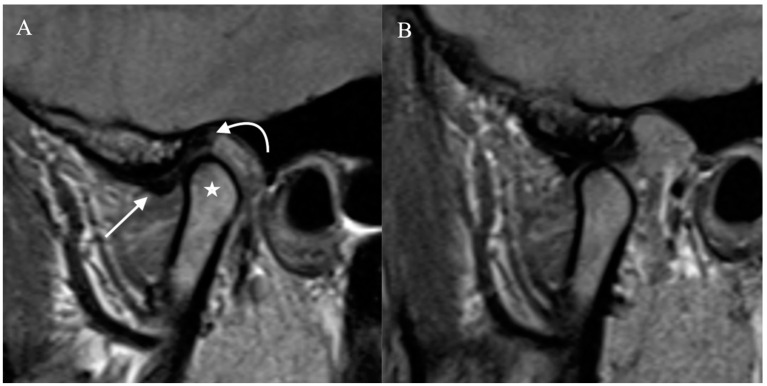
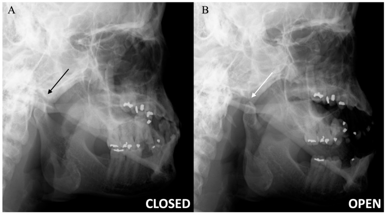
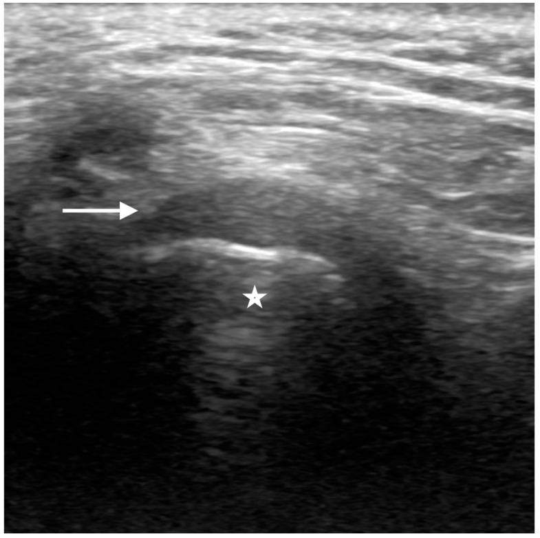
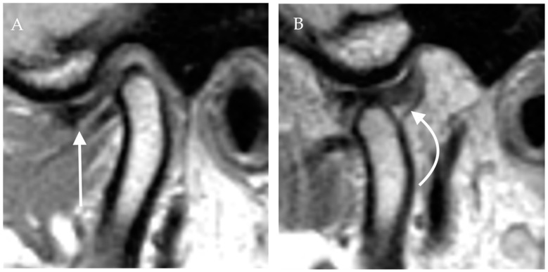
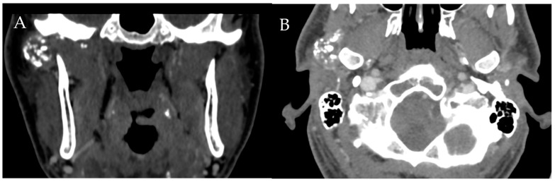
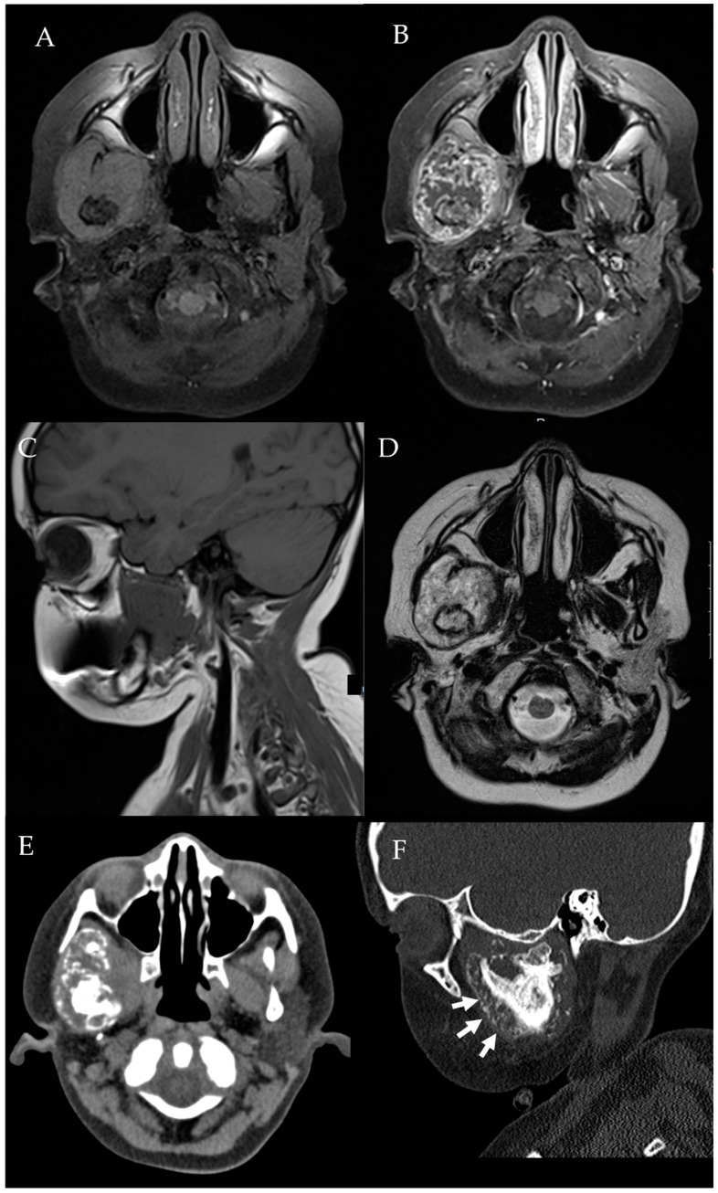
Temporomandibular disorder (TMD) is a common musculoskeletal condition that causes pain and disability for patients and imposes a high financial burden on the healthcare system. The most common cause of TMD is internal derangement, mainly secondary to articular disc displacement. Multiple other pathologies such as inflammatory arthritis, infection, and neoplasm can mimic internal derangement. MRI is the modality of choice for evaluation of the TMJ. Radiologists need to be familiar with the normal anatomy and function of the TMJ and MR imaging of the internal derangement and other less common pathologies of the TMJ.
Keywords: TMJ; arthritis; imaging; internal derangement; neoplasm; radiologist; temporomandibular disorder; temporomandibular joint.
Conflict of interest statement
The authors declare that they have no conflict of interest.
Figures

References
-
- Slade G.D., Fillingim R.B., Sanders A.E., Bair E., Greenspan J.D., Ohrbach R., Dubner R., Diatchenko L., Smith S.B., Knott C., et al. Summary of Findings from the OPPERA Prospective Cohort Study of Incidence of First-Onset Temporomandibular Disorder: Implications and Future Directions. J. Pain. 2013;14:T116–T124. doi: 10.1016/j.jpain.2013.09.010. - DOI - PMC - PubMed
Publication types
LinkOut - more resources
Full Text Sources
