. 2022 Apr 18;12(4):1017.
doi: 10.3390/diagnostics12041017.

Novel User-Friendly Application for MRI Segmentation of Brain Resection following Epilepsy Surgery

Affiliations

Novel User-Friendly Application for MRI Segmentation of Brain Resection following Epilepsy Surgery

Roberto Billardello et al. Diagnostics (Basel). .

Abstract

Delineation of resected brain cavities on magnetic resonance images (MRIs) of epilepsy surgery patients is essential for neuroimaging/neurophysiology studies investigating biomarkers of the epileptogenic zone. The gold standard to delineate the resection on MRI remains manual slice-by-slice tracing by experts. Here, we proposed and validated a semiautomated MRI segmentation pipeline, generating an accurate model of the resection and its anatomical labeling, and developed a graphical user interface (GUI) for user-friendly usage. We retrieved pre- and postoperative MRIs from 35 patients who had focal epilepsy surgery, implemented a region-growing algorithm to delineate the resection on postoperative MRIs and tested its performance while varying different tuning parameters. Similarity between our output and hand-drawn gold standards was evaluated via dice similarity coefficient (DSC; range: 0-1). Additionally, the best segmentation pipeline was trained to provide an automated anatomical report of the resection (based on presurgical brain atlas). We found that the best-performing set of parameters presented DSC of 0.83 (0.72-0.85), high robustness to seed-selection variability and anatomical accuracy of 90% to the clinical postoperative MRI report. We presented a novel user-friendly open-source GUI that implements a semiautomated segmentation pipeline specifically optimized to generate resection models and their anatomical reports from epilepsy surgery patients, while minimizing user interaction.

Keywords: MRI; brain resection; epilepsy surgery; image segmentation; region growing.


Conflict of interest statement

The authors declare no conflict of interest.

Figures
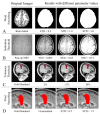
Figure 1
Figure 1
Results from low (2nd column) to high (4th column) parameter values. Mask Smoothing (A-1st row): original preoperative MRI brain mask (left) and three different binarized smoothed masks (right); Saturation Threshold (B-2nd row): low-contrast original postoperative MRI (left) and three contrast-adjusted images (right); Tolerance Threshold (C-3rd row): manual traced resection (left) and three examples of resections obtained with increasing tolerance value (right); and Final Smoothing (D-4th row): manual traced resection (left) and three different values of smoothing.
Figure 2
Figure 2
Seed positioning in different MRI views. We selected four seeds (indicated by red stars) from different slices from each MRI view (first row: axial view, second row: coronal view, third row: sagittal view), obtaining a total of 12 different seeds for each patient.
Figure 3
Figure 3
Automated generation of the resection anatomical report. Example of inputs (right: segmentation of the resection cavity, preoperative Desikan–Killiany cortical parcellation) and outputs (left: resection percentages of each anatomical area, and list of resected areas). Each cortical area of the Desikan–Killiany is associated with one anatomical area (frontal, temporal, parietal, occipital, cingulate, hippocampus, amygdala). Two approaches (overlap- and KNN-based) were used to estimate the resection percentage of each anatomical area and then generate an anatomical report, which was then validated by comparison with the ground truth (clinical MRI postsurgical report). In this example, both automated reports indicate right parietal lobe resection, consistently with the ground truth.
Figure 4
Figure 4
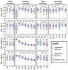
DSC changes using different parameter values. Each boxplot shows the DSC of all the pipelines where one parameter was set to a certain value. Significant increases and decreases are indicated with a star and a triangle. Boxbplots corresponding to the optimal values are in green. Top edges of the boxes indicate the 25th and 75th percentiles.
Figure 5
Figure 5
DSC for the best-performing pipeline of each standardization method. Pies indicate the percentage of patients with excellent similarity (yellow, DSC > 0.8%), high similarity (green, DSC from 0.7 to 0.8), good similarity (blue, DSC from 0.6 to 0.7), and poor similarity (DSC < 0.6). Significant differences (Wilcoxon signed-rank test) between the standardization methods are indicated with asterisks (* p < 0.05; ** p < 0.01). Data points beyond the boxplot whiskers are indicated with +.
Figure 6
Figure 6
Anatomical report results. Overall performances (left bar plot) and individual reports’ concordance (right bar plot) for overlap-based (left) and KNN-label-substitution (right) approaches.
Figure 7
Figure 7
GUI for the resection segmentation. (A): Interface to select postoperative MRI and preoperative brain-mask files, or the patient’s Brainstorm anatomy folder that contains them. (B): Slider to select tolerance threshold (default: optimal value). (C): Interface to place the initial seed using an MRI viewer (see Figure 8) or entering its coordinates. The user can set an additional ROI for region growing by entering its ranges (this is optional, see Figure 8). (D): Options to exclude brain ventricles and/or generate the anatomical report through a brain parcellation file, which must be selected. E and F: the user can change the view (E) and slice (F) to visualize the postoperative MRI and resection model shown in G. (G): Preview of results (left: postoperative MRI; middle: resection model, right: automated report and resection volume in cm3). (H): Buttons to run the segmentation (“Calculate”), or to export results to a file (NIfTI), MATLAB workspace, or directly to the initial Brainstorm anatomy folder. Status LED turns red in case of error (green otherwise). “Show 3D” button shows the resection model (see Figure 8), while “Reset” allows to reset all settings and run again.
Figure 8
Figure 8
GUI popup windows. (A) MRI viewer for seed placement (when “Place Manually” is clicked). (B) Window for the definition of an optional ROI in which the region is free to grow (when “Change Ranges” button is clicked). (C) 3D MRI viewer from Brainstorm software (if “Show 3D” button is clicked) shows the 3D resection output (red). (D) If “Export to Workspace” is clicked, the user can export the output in voxel coordinates and/or in Subject Coordinates System (SCS), as well as the segmentation parameters and seed coordinates (Segmentation info). (E) Bar graph of the anatomical report (when “Generate automated report graphs” is selected).
Figure 9
Figure 9
Examples of segmentation from the presented GUI from three different patients: left frontal resection (#1), left temporal resection (#2); right parietal resection (#3).

References

    1. Vakharia V.N., Duncan J.S., Witt J.-A., Elger C.E., Staba R., Engel J. Getting the Best Outcomes from Epilepsy Surgery. Ann. Neurol. 2018;83:676–690. doi: 10.1002/ana.25205. - DOI - PMC - PubMed
    1. Tamilia E., Park E.H., Percivati S., Bolton J., Taffoni F., Peters J.M., Grant P.E., Pearl P.L., Madsen J.R., Papadelis C. Surgical Resection of Ripple Onset Predicts Outcome in Pediatric Epilepsy. Ann. Neurol. 2018;84:331–346. doi: 10.1002/ana.25295. - DOI - PubMed
    1. Ryvlin P., Cross J.H., Rheims S. Epilepsy Surgery in Children and Adults. Lancet Neurol. 2014;13:1114–1126. doi: 10.1016/S1474-4422(14)70156-5. - DOI - PubMed
    1. Abdallah C., Maillard L.G., Rikir E., Jonas J., Thiriaux A., Gavaret M., Bartolomei F., Colnat-Coulbois S., Vignal J.P., Koessler L. Localizing Value of Electrical Source Imaging: Frontal Lobe, Malformations of Cortical Development and Negative MRI Related Epilepsies Are the Best Candidates. NeuroImage Clin. 2017;16:319–329. doi: 10.1016/j.nicl.2017.08.009. - DOI - PMC - PubMed
    1. Rosenow F., Lüders H. Presurgical Evaluation of Epilepsy. Brain. 2001;124:1683–1700. doi: 10.1093/brain/124.9.1683. - DOI - PubMed

LinkOut - more resources