Review
. 2022 Mar 31;12(4):530.
doi: 10.3390/biom12040530.

Cellular Distribution of Brain Aquaporins and Their Contribution to Cerebrospinal Fluid Homeostasis and Hydrocephalus

Affiliations
Review

Cellular Distribution of Brain Aquaporins and Their Contribution to Cerebrospinal Fluid Homeostasis and Hydrocephalus

José Luis Trillo-Contreras et al. Biomolecules. .

Abstract

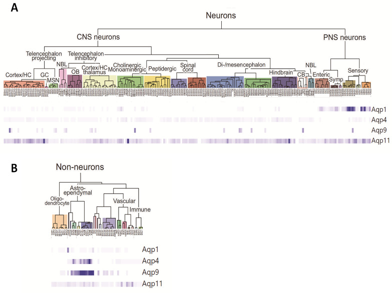
Brain aquaporins facilitate the movement of water between the four water compartments: blood, cerebrospinal fluid, interstitial fluid, and intracellular fluid. This work analyzes the expression of the four most abundant aquaporins (AQPs) (AQP1, AQP4, AQP9, and AQP11) in the brains of mice and discuss their contribution to hydrocephalus. We analyzed available data from single-cell RNA sequencing of the central nervous system of mice to describe the expression of aquaporins and compare their distribution with that based on qPCR, western blot, and immunohistochemistry assays. Expression of AQP1 in the apical cell membrane of choroid plexus epithelial cells and of AQP4 in ependymal cells, glia limitans, and astrocyte processes in the pericapillary end foot is consistent with the involvement of both proteins in cerebrospinal fluid homeostasis. The expression of both aquaporins compensates for experimentally induced hydrocephalus in the animals. Recent data demonstrate that hypoxia in aged animals alters AQP4 expression in the choroidal plexus and cortex, increasing the ventricle size and intraventricular pressure. Cerebral distensibility is reduced in parallel with a reduction in cerebrospinal fluid drainage and cognitive deterioration. We propose that aged mice chronically exposed to hypoxia represent an excellent experimental model for studying the pathophysiological characteristics of idiopathic normal pressure hydrocephalus and roles for AQPs in such disease.

Keywords: aging; aquaporins; cerebrospinal fluid; choroid plexus; ependyma; hydrocephalus; hypoxia; sequencing; single-cell RNA.


Conflict of interest statement

The authors declare no conflict of interest.

Figures

Figure 1
Figure 1
Phylogenetic tree of human aquaporins and other known aquaporins. The 13 aquaporins known to date in humans are represented, according to the homology in their amino acid sequence for each member of the family, differentiating between “strict” aquaporins (water aquaporins, at the bottom) and aquaglyceroporins (at the top). Superaquaporins, the last aquaporins identified in mammals are also shown and are the most distant in their amino acid sequence homology from the two groups mentioned above but are closer to the aquaporins expressed in species of intracellular parasites such as Trypanosoma cruzi (TcAQP) and of nematodes such as Caenorhabditis elegans and Caenorhabditis briggsae. The tree also shows certain bacterial aquaporins such as GlpF, a facilitator of the passage of glycerol, which are similar in their sequence to the aquaglyceroporins, and AQPZ from E. coli, as well as the aquaporin from methanogen archaea, AQPM from Methanothermobacter marburgensis and the plant aquaporins from Arabidopsis thaliana (small basic intrinsic protein [SIP] and plasma membrane intrinsic protein [PIP], respectively). Modified from Gorelick et al., 2006 [17].
Figure 2
Figure 2
Molecular study of the mouse nervous system using single-cell RNA sequencing technology. The 265 distinct cell clusters identified based on the analysis of 160,796 transcriptomes of the mouse nervous system: brain, spinal cord, dorsal and peripheral sympathetic ganglia, and enteric nervous system are represented and classified in the tree diagrams; (A) Tree diagram for Neuron clusters, and (B) Tree diagram for non-neuron clusters. The represented dendrogram has been constructed using the digital resource (http://mousebrain.org; accessed on 23 October 2019); prepared in the laboratory of Sten Linnarsson, obtained from Zeisel et al. (2018) [81], and shows the relationship between cell types and the gene expression of AQP1, AQP4, AQP9, and AQP11. The color-graded bands at the bottom of the figure show the expression pattern of each gene in the 265 identified cell types. Each band represents the relative expression of a transcript in a cell type with respect to the maximum expression detected for that same transcript in any cell type. Given that they are relative data, the richness of expression of the different transcripts cannot be compared with each other, but they do help locate where the expression occurs. Thus, AQP4 and AQP9 appear to show a very similar expression pattern, clearly predominant in the astroependymal line, although their expression levels are very different. AQP1 expression is well defined in the dorsal ganglia and ependymal cells of the choroid plexus and contrasts with the high delocalization of AQP11. The data on absolute expression are represented in Table 2.
Figure 3
Figure 3
AQP1 and AQP4 expression in the choroid plexus of mice exposed to hypoxia: (A) The AQP mRNA expression in the choroid plexus (Plx) of mice exposed to hypoxia (10% O2, 48 h) and normoxic controls was analyzed by qPCR. 18S ribosomal RNA was used as the housekeeping gene for normalization (modified from Trillo et al., 2018 [115]). (B) AQP1 and AQP4 immunofluorescence images of the choroid plexus obtained from mice exposed to normoxia or hypoxia (10% O2; 48 h). AQP4 expression is notably higher after the hypoxic treatment. (C) Quantification by optical density of AQP1 and AQP4 expression in the choroid plexus tissue (Modified from Trillo et al., 2018 [115]) (* p < 0.05; *** p < 0.001).

References

    1. Dix J.A., Solomon A. Role of membrane proteins and lipids in water diffusion across red cell membranes. Biochim. Biophys. Acta (BBA)-Biomembr. 1984;773:219–230. doi: 10.1016/0005-2736(84)90085-3. - DOI - PubMed
    1. Whittembury G., Carpi-Medina P., Gonzalez E., Linares H. Effect of para-chloromercuribenzenesulfonic acid and temperature on cell water osmotic permeability of proximal straight tubules. Biochim. Biophys. Acta (BBA)-Biomembr. 1984;775:365–373. doi: 10.1016/0005-2736(84)90192-5. - DOI - PubMed
    1. Brown P.A., Feinstein M.B., Sha’Afi R.I. Membrane proteins related to water transport in human erythrocytes. Nature. 1975;254:523–525. doi: 10.1038/254523a0. - DOI - PubMed
    1. Meyer M.M., Verkman A.S. Evidence for water channels in renal proximal tubule cell membranes. J. Membr. Biol. 1987;96:107–119. doi: 10.1007/BF01869237. - DOI - PubMed
    1. Echevarria M., González E., Gutierrez A., Whittembury G. Water and urea diffusive permeabilities in isolated proximal tubule cells. Am. J. Physiol. Physiol. 1994;267:F709–F715. doi: 10.1152/ajprenal.1994.267.5.F709. - DOI - PubMed

Publication types