Review
. 2022 Apr 7;14(8):1860.
doi: 10.3390/cancers14081860.

Real-Time MRI-Guided Prostate Interventions

Affiliations
Review

Real-Time MRI-Guided Prostate Interventions

Seyedeh Nina Masoom et al. Cancers (Basel). .

Abstract

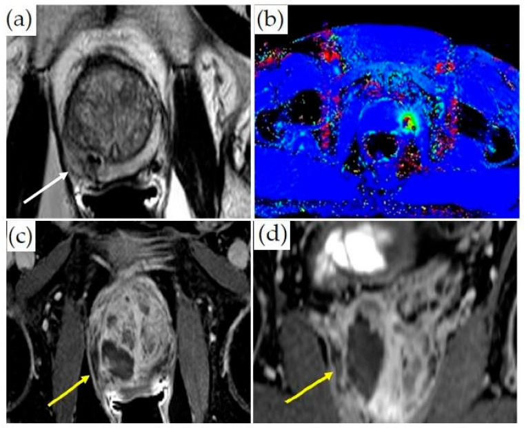
Prostate cancer (PCa) is the second most common cause of cancer death in males. Targeting MRI-visible lesions has led to an overall increase in the detection of clinically significant PCa compared to the prior practice of random ultrasound-guided biopsy of the prostate. Additionally, advances in MRI-guided minimally invasive focal treatments are providing new options for patients with PCa. This review summarizes the currently utilized real-time MRI-guided interventions for PCa diagnosis and treatment.

Keywords: MRI-guided prostate interventions; multiparametric magnetic resonance imaging (mpMRI); prostate ablation; prostate biopsy; prostate cancer (PCa).


Conflict of interest statement

Author P.G. is on the Medical Advisory Boards of INSIGHTEC and SonoALASense and has an ownership/equity interest in SonoALASense. Author A.O. is the co-founder and co-owner of QMIS; he is on the Medical Advisory Board of Profound and has research grants from Profound and Philips. Author S.A. is involved with a clinical trial and has research support from Profound. The remaining authors declare no conflict of interest.

Figures

Figure 1
Figure 1
In-bore transperineal biopsy: (a) diagram of a patient placed supine in an MRI-compatible stirrups device and grid; (b) coronal and (c) sagittal T1-weighted MRI images demonstrating the needle entering the right prostate gland (white arrows).
Figure 2
Figure 2
An office-based, low-field MRI system for prostate biopsy: (a) low-field MRI device; (b) biopsy device; (c) axial T2-weighted images acquired on the low-field magnet; (d) registered high-resolution axial T2-weighted image and grid for biopsy planning. A target is identified in the left lateral prostate (white arrow).
Figure 3
Figure 3
Treatment of a patient with localized, low-stage PCa with a TULSA-PRO device: (a) Sagittal T2-weighted MR image demonstrates the transurethral sonoablation device within the prostatic urethra and the tip within the bladder of a patient without a rectum (white arrow). (b) Axial T2-weighted MR planning images of the prostate at the level of 8 ultrasound transducers. The prostate boundary is contoured in orange. (c) MR thermometry images and color overlay demonstrate areas of heating. (d) Post-treatment imaging of the prostate acquired after gadolinium administration. Areas of bright signal demonstrate viable tissue, while dark areas indicate treated tissue.
Figure 4
Figure 4
Treatment of a patient with Gleason 3 + 4 PCa with transrectal MR-guided HIFU (ExAblate 2100 device): (a) Pretreatment axial T2-weighted MR imaging demonstrates a hypointense lesion in the medial left peripheral zone (white arrow). (b) Overlayed treatment planning image shows the prostate contour (blue contour), region of interest with at least 5 mm margins (orange contour), focal spots (green dots inside rectangles), predicted thermal dose (green contour), and lesion (white arrow). (c) Thermal map overlay image demonstrates areas that have reached thermal dose based on intraoperative thermometry (dark blue at least 8000 CEM43, light blue 240 CEM43). (d) Axial post-contrast images obtained immediately after treatment demonstrate a non-perfused area (dark area, yellow arrow) indicating treatment. The viable prostate tissue demonstrates enhancement (bright signal, asterisk).
Figure 5
Figure 5
MRI-guided laser ablation: (a) Axial T2-weighted MR images with lesion in the right medial peripheral zone (white arrow). (b) Thermometry mapping (different patient) during treatment. (c) Axial and (d) coronal post-contrast T1-weighted imaging demonstrating ablation cavity (yellow arrows).
Figure 6
Figure 6
MRI-guided cryoablation and monitoring of ice-ball formation on axial T1-weighted fat-saturated images: (ac) Ice-ball formation as a function of time in the right lateral peripheral zone of the prostate (white arrowheads). The untreated normal prostate tissue is visualized.

References

    1. GLOBOCAN World Health Organization. [(accessed on 2 February 2022)]. Available online: https://gco.iarc.fr/today/data/factsheets/populations/900-world-fact-she....
    1. Cooperberg M.R., Broering J.M., Kantoff P.W., Carroll P.R. Contemporary trends in low risk prostate cancer: Risk assessment and treatment. J. Urol. 2007;178:s14–s19. doi: 10.1016/j.juro.2007.03.135. - DOI - PMC - PubMed
    1. Quon H., Loblaw A., Nam R. Dramatic increase in prostate cancer cases by 2021. BJU Int. 2011;108:1734–1738. doi: 10.1111/j.1464-410X.2011.10197.x. - DOI - PubMed
    1. Moyer V.A., U.S. Preventive Services Task Force Screening for prostate cancer: U.S. Preventive Services Task Force recommendation statement. Ann. Intern. Med. 2012;157:120–134. doi: 10.7326/0003-4819-157-2-201207170-00459. - DOI - PubMed
    1. Schröder F.H., Hugosson J., Roobol M.J., Tammela T.L., Zappa M., Nelen V., Kwiatkowski M., Lujan M., Määttänen L., Lilja H., et al. ERSPC Investigators. Screening and prostate cancer mortality: Results of the European Randomised Study of Screening for Prostate Cancer (ERSPC) at 13 years of follow-up. Lancet. 2014;384:2027–2035. doi: 10.1016/S0140-6736(14)60525-0. - DOI - PMC - PubMed

LinkOut - more resources