Review
. 2022 Apr 7;14(8):1865.
doi: 10.3390/cancers14081865.

Extracellular Vesicle-Mediated Mitochondrial Reprogramming in Cancer

Affiliations
Review

Extracellular Vesicle-Mediated Mitochondrial Reprogramming in Cancer

Roger Carles-Fontana et al. Cancers (Basel). .

Abstract

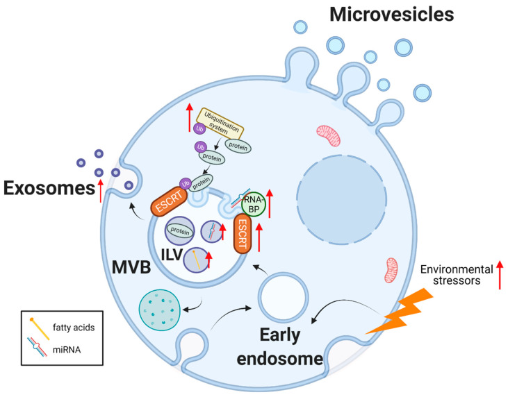
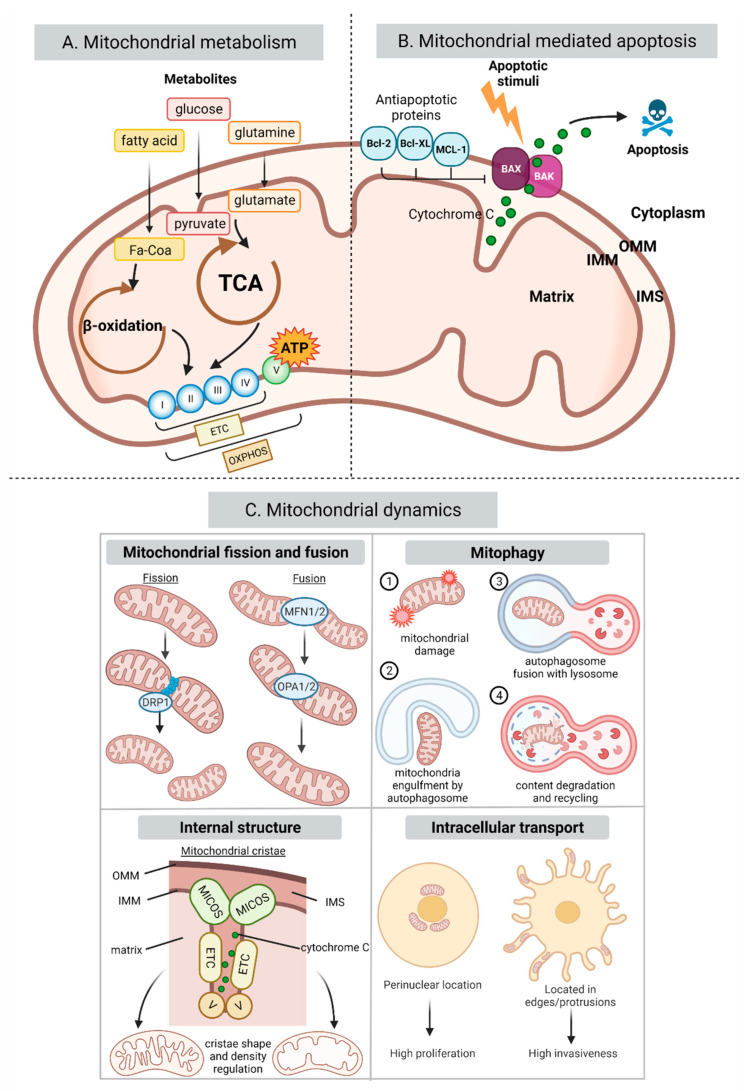
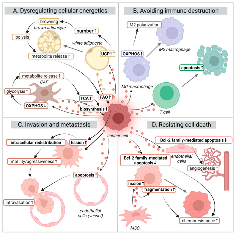
Altered metabolism is a defining hallmark of cancer. Metabolic adaptations are often linked to a reprogramming of the mitochondria due to the importance of these organelles in energy production and biosynthesis. Cancer cells present heterogeneous metabolic phenotypes that can be modulated by signals originating from the tumor microenvironment. Extracellular vesicles (EVs) are recognized as key players in intercellular communications and mediate many of the hallmarks of cancer via the delivery of their diverse biological cargo molecules. Firstly, this review introduces the most characteristic changes that the EV-biogenesis machinery and mitochondria undergo in the context of cancer. Then, it focuses on the EV-driven processes which alter mitochondrial structure, composition, and function to provide a survival advantage to cancer cells in the context of the hallmarks of cancers, such as altered metabolic strategies, migration and invasiveness, immune surveillance escape, and evasion of apoptosis. Finally, it explores the as yet untapped potential of targeting mitochondria using EVs as delivery vectors as a promising cancer therapeutic strategy.

Keywords: metabolism; miRNA; mitochondrial dynamics; tumor microenvironment (TME); tumor-derived EVs (TEVs).


Conflict of interest statement

The authors declare no conflict of interest.

Figures

Figure 1
Figure 1
EV biogenesis, release, and cargo in cancer. Exosome biogenesis starts with the inward budding of the plasma membrane (early endosome). The limiting membrane buds inwards, generating intraluminal vesicles (ILV) contained in a late endosome or multivesicular body (MVB). The membrane of the MVB body fuses with the plasma membrane releasing the exosomes to the extracellular milieu. Several of the elements involved in exosome biogenesis and cargo sorting are upregulated in cancer (marked with a red arrow). Microvesicles or ectosomes are generated by the direct outward budding of the plasma membrane. Abbreviations used: ESCRT: endosomal sorting complexes required for transport. RNA-BP: RNA binding proteins. Ub: ubiquitin. miRNA: microRNA. Figure created with BioRender.com (accessed on 31 March 2022).
Figure 2
Figure 2
Mitochondrial processes affected in cancer. (A) Summarizes key metabolic processes in mitochondria. Metabolites such as glucose, fatty acids (FA) and glutamine are transformed into pyruvate, FA-CoA and glutamate and enter the TCA and β-oxidation cycles taking place in the mitochondrial matrix. These processes are used by the ETC to pump protons into the intermembrane space creating a chemical gradient used by complex V to generate ATP in a process called OXPHOS. (B) Summarizes the role of the Bcl-2 family proteins in the mitochondrial intrinsic pathway. Briefly, apoptotic stimuli lead to the formation of the pores in the OMM by Bax and Bak releasing cytochrome c into the cytoplasm which triggers the caspase cascade inducing apoptosis. The antiapoptotic members of the Bcl-2 family inhibit this process. (C) Summarizes the four main mitochondrial dynamics processes: mitochondrial fusion and fission, mitophagy, internal cristae architecture, and mitochondrial intracellular trafficking. In cancer, all these mitochondrial activities are dysregulated to support cancer energetic and biosynthetic demands, as well as survival. Abbreviations used: TCA: tricarboxylic acid. ATP: adenosine triphosphate. ETC: electron transport chain. OXPHOS: oxidative phosphorylation. I: complex I. II: complex II. III: complex III. IV: complex IV. V: complex V. OMM: outer mitochondrial membrane. IMM: inner mitochondrial membrane. IMS: intermembrane space. MICOS: mitochondrial contact site and cristae organizing system. Figure created with BioRender.com (accessed on 31 March 2022).
Figure 3
Figure 3
EV modulation of mitochondria and its effects in the context of hallmarks of cancer. Cancer cells generate tumor-derived EVs (TEVs) that alter the mitochondrial behavior of different cell types of the tumor microenvironment to support cancer progression, invasion, and survival. Through these effects, TEVs are mediators of several of the hallmarks of cancer such as: (A) dysregulation of cellular energetics: cancer cells can induce the release of metabolites from CAFs and adipocytes to be used for cancer cell biosynthesis and FAO; (B) avoiding immune destruction: TEVs can induce immunosuppression by inducing M2 polarization in macrophages or T cell apoptosis via the mitochondrial intrinsic pathway; (C) promoting cell motility and facilitating invasiveness: increased FAO leads to an intracellular trafficking of mitochondria towards edge protrusions facilitating their migration. At the same time, they can induce mitochondrial apoptosis in endothelial barrier cells to permit intravasation; (D) resisting cell death: alterations in Bcl-2 family members leads to inhibition of mitochondrial apoptosis resulting in chemotherapy resistance and angiogenesis. Abbreviations used: OXPHOS: oxidative phosphorylation. TCA: tricarboxylic acid cycle. FAO: fatty acid metabolism. UCP1: uncoupling protein 1. MSC: mesenchymal stem cell. Labels in bold represent mitochondria-related elements and processes. The colors of the EVs reflect those of the parental cell that produced them. The colors of the text borders reflect the cells where the indicated processes take place. Figure created with BioRender.com (accessed on 31 March 2022).

References

    1. Hanahan D., Weinberg R.A. Hallmarks of cancer: The next generation. Cell. 2011;144:646–674. doi: 10.1016/j.cell.2011.02.013. - DOI - PubMed
    1. Cassim S., Pouyssegur J. Tumor Microenvironment: A Metabolic Player that Shapes the Immune Response. Int. J. Mol. Sci. 2019;21:157. doi: 10.3390/ijms21010157. - DOI - PMC - PubMed
    1. Bebelman M., Smit M.J., Pegtel D.M., Baglio S.R. Biogenesis and function of extracellular vesicles in cancer. Pharmacol. Ther. 2018;188:1–11. doi: 10.1016/j.pharmthera.2018.02.013. - DOI - PubMed
    1. Théry C., Witwer K.W., Aikawa E., Alcaraz M.J., Anderson J.D., Andriantsitohaina R., Antoniou A., Arab T., Archer F., Atkin-Smith G.K., et al. Minimal information for studies of extracellular vesicles 2018 (MISEV2018): A position statement of the International Society for Extracellular Vesicles and update of the MISEV2014 guidelines. J. Extracell. Vesicles. 2018;7:1535750. doi: 10.1080/20013078.2018.1535750. - DOI - PMC - PubMed
    1. Maisano D., Mimmi S., Dattilo V., Marino F., Gentile M., Vecchio E., Fiume G., Nisticò N., Aloisio A., de Santo M.P., et al. A novel phage display based platform for exosome diversity characterization. Nanoscale. 2022;14:2998–3003. doi: 10.1039/D1NR06804K. - DOI - PubMed

LinkOut - more resources