. 2022 Apr 8;14(8):1890.
doi: 10.3390/cancers14081890.

Deciphering HER2-HER3 Dimerization at the Single CTC Level: A Microfluidic Approach

Affiliations

Deciphering HER2-HER3 Dimerization at the Single CTC Level: A Microfluidic Approach

Ezgi Tulukcuoglu Guneri et al. Cancers (Basel). .

Abstract

Microfluidics has provided clinicians with new technologies to detect and analyze circulating tumor biomarkers in order to further improve their understanding of disease mechanism, as well as to improve patient management. Among these different biomarkers, circulating tumor cells have proven to be of high interest for different types of cancer and in particular for breast cancer. Here we focus our attention on a breast cancer subtype referred as HER2-positive breast cancer, this cancer being associated with an amplification of HER2 protein at the plasma membrane of cancer cells. Combined with therapies targeting the HER2 protein, HER2-HER3 dimerization blockade further improves a patient's outcome. In this work, we propose a new approach to CTC characterization by on-chip integrating proximity ligation assay, so that we can quantify the HER2-HER3 dimerization event at the level of single CTC. To achieve this, we developed a microfluidic approach combining both CTC capture, identification and HER2-HER3 status quantification by Proximity Ligation Assay (PLA). We first optimized and demonstrated the potential of the on-chip quantification of HER2-HER3 dimerization using cancer cell lines with various levels of HER2 overexpression and validated its clinical potential with a patient's sample treated or not with HER2-targeted therapy.

Keywords: HER2; HER3 dimerization; circulating tumor cells; microfluidic; proximity ligation assay.


Conflict of interest statement

The authors declare no conflict of interest.

Figures

Figure 1
Figure 1
(a) Schematic representation of indirect PLA; primary antibodies are detected by PLA conjugated secondary antibodies. (b) Direct PLA depicted by conjugated primary antibodies only. (c) Direct PLA signals detected in SK-BR-3 cells in the presence of HER2 and HER3 antibodies (left). No signals detected after omitting HER3 antibody (right). (d) Schematic representation of the direct PLA-Assay. (e) PLA coupled with immunostaining with SK-BR-3 cells on glass slides. Scale bar is 20 µm (c,d), 10 µm (e). Schemes were created by using Servier Medical Art, 2021.
Figure 2
Figure 2
(a) Picture of the COC Ephesia chip. (b) Schematic representation of Ephesia CTC immunoextraction; captured cells in the array of self-assembled anti-EpCAM beads are subjected to on-chip PLA. B arrow shows the magnetic field direction. (c) SK-BR-3 cells captured inside Ephesia device showing PLA signals (orange) and stained for cytokeratin CK (green), CD45 (red) and DAPI (blue). Scale bar is 10 µm. (Schemes were created by using Servier Medical Art, 2021.
Figure 3
Figure 3
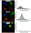
Left panel: On-chip immunofluorescence images of (a) MDA-MB-231 cells and (b) SK-BR-3 cells captured inside Ephesia showing PLA signals (left panel, orange). (c) HER2/HER3 gene silencing in SK-BR-3 cells induced a decreased in PLA signals, represented by the blue Gaussian curve shifted to the left. Right panel: Graphs quantifying the dimerization events per cell (bottom right, red) in each condition. n = 3 independent experiments for each condition. Scale bar: 10 µm.
Figure 4
Figure 4
(a) Examples of CTCs detected from patient blood with MBC HER2− or HER2+, using the Ephesia chip. CTCs were further subjected to PLA and immunostaining for CK, DAPI and CD45 detection. (b) Violin plots showing the distribution of HER2-HER3 PLA signal per cell (number of CTC detected indicated for each patient sample). Scale bar is 10 µm.

References

    1. Perou C.M., Sørlie T., Eisen M.B., van de Rijn M., Jeffrey S.S., Rees C.A., Pollack J.R., Ross D.T., Johnsen H., Akslen L.A., et al. Molecular portraits of human breast tumours. Nature. 2000;406:747–752. doi: 10.1038/35021093. - DOI - PubMed
    1. Goutsouliak K., Veeraraghavan J., Sethunath V., De Angelis C., Osborne C.K., Rimawi M.F., Schiff R. Towards personalized treatment for early stage HER2-positive breast cancer. Nat. Rev. Clin. Oncol. 2019;17:233–250. doi: 10.1038/s41571-019-0299-9. - DOI - PMC - PubMed
    1. Wolff A.C., Hammond M.E.H., Hicks D.G., Dowsett M., McShane L.M., Allison K.H., Allred D.C., Bartlett J.M., Bilous M., Fitzgibbons P., et al. Recommendations for human epidermal growth factor receptor 2 testing in breast cancer: American Society of Clinical Oncology/College of American Pathologists clinical practice guideline update. J. Clin. Oncol. 2013;31:3997–4013. doi: 10.1200/JCO.2013.50.9984. - DOI - PubMed
    1. Yamashita-Kashima Y., Shu S., Yorozu K., Moriya Y., Harada N. Mode of action of pertuzumab in combination with trastuzumab plus docetaxel therapy in a HER2-positive breast cancer xenograft model. Oncol. Lett. 2017;14:4197–4205. doi: 10.3892/ol.2017.6679. - DOI - PMC - PubMed
    1. Sawyers C.L. Herceptin: A fist assault on oncongenes that launched a revolution. Cell. 2019;197:8–12. doi: 10.1016/j.cell.2019.08.027. - DOI - PubMed