. 2022 Apr 9;14(8):1900.
doi: 10.3390/cancers14081900.

Aspirin-Triggered Resolvin D1 Reduces Chronic Dust-Induced Lung Pathology without Altering Susceptibility to Dust-Enhanced Carcinogenesis

Affiliations

Aspirin-Triggered Resolvin D1 Reduces Chronic Dust-Induced Lung Pathology without Altering Susceptibility to Dust-Enhanced Carcinogenesis

Edward C Dominguez et al. Cancers (Basel). .

Abstract

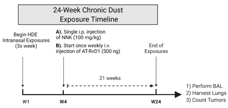
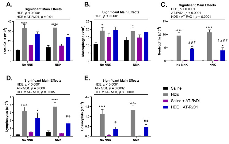
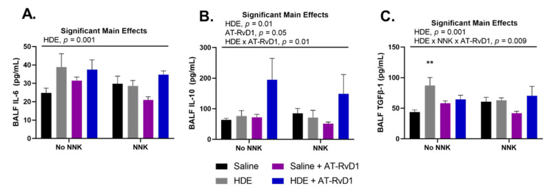
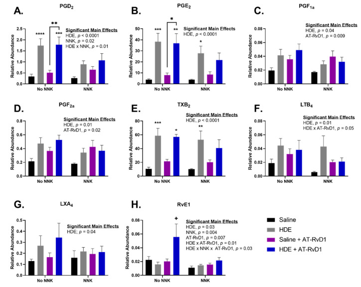
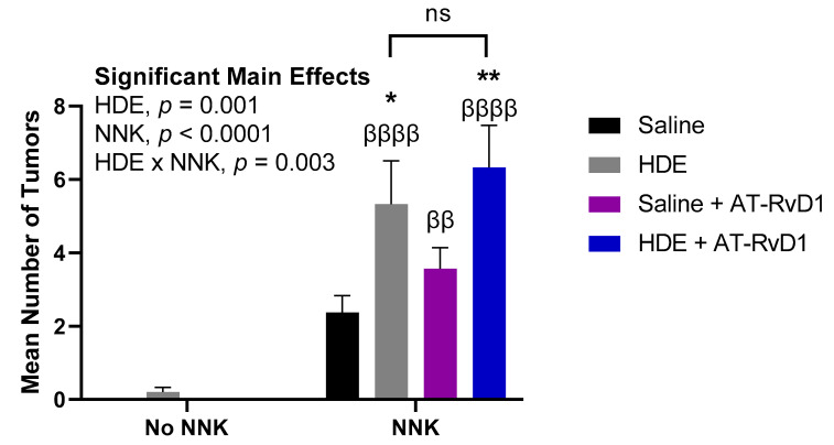
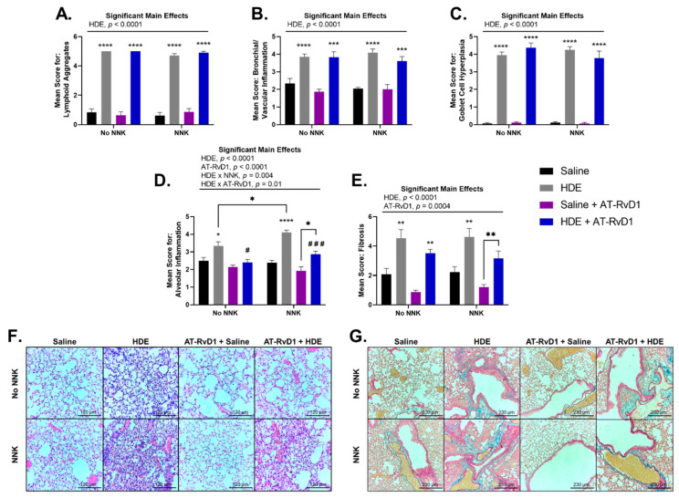
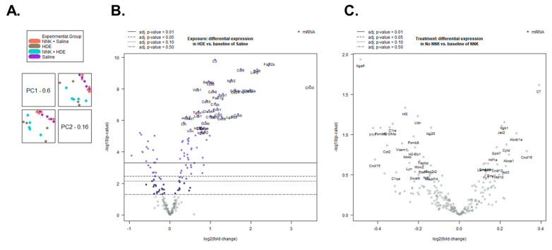
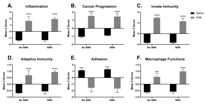
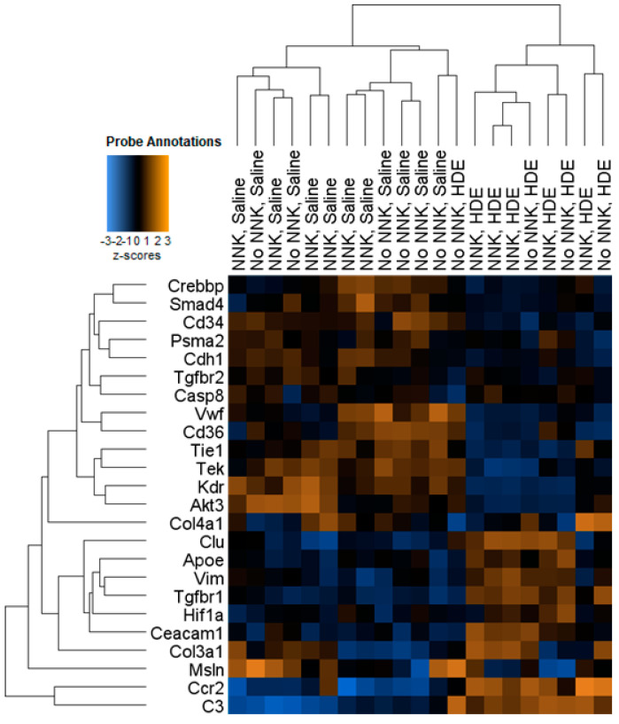
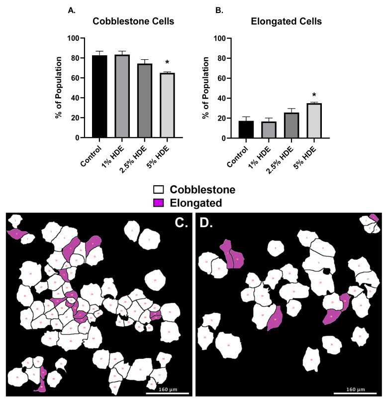
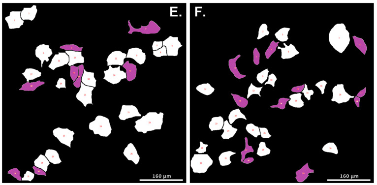
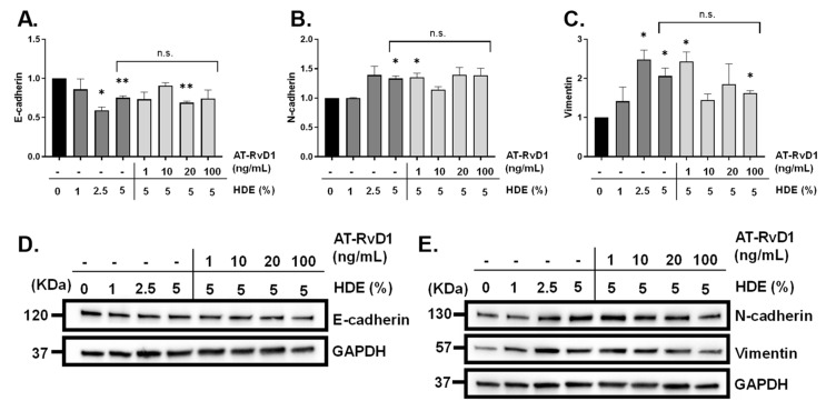
Lung cancer is the leading cause of cancer-related deaths worldwide, with increased risk being associated with unresolved or chronic inflammation. Agricultural and livestock workers endure significant exposure to agricultural dusts on a routine basis; however, the chronic inflammatory and carcinogenic effects of these dust exposure is unclear. We have developed a chronic dust exposure model of lung carcinogenesis in which mice were intranasally challenged three times a week for 24 weeks, using an aqueous dust extract (HDE) made from dust collected in swine confinement facilities. We also treated mice with the omega-3-fatty acid lipid mediator, aspirin-triggered resolvin D1 (AT-RvD1) to provide a novel therapeutic strategy for mitigating the inflammatory and carcinogenic effects of HDE. Exposure to HDE resulted in significant immune cell influx into the lungs, enhanced lung tumorigenesis, severe tissue pathogenesis, and a pro-inflammatory and carcinogenic gene signature, relative to saline-exposed mice. AT-RvD1 treatment mitigated the dust-induced inflammatory response but did not protect against HDE + NNK-enhanced tumorigenesis. Our data suggest that chronic HDE exposure induces a significant inflammatory and pro-carcinogenic response, whereas treatment with AT-RvD1 dampens the inflammatory responses, providing a strong argument for the therapeutic use of AT-RvD1 to mitigate chronic inflammation.

Keywords: epithelial to mesenchymal transition (EMT); fibrosis; lung cancer; lung inflammation; omega-3 fatty acids; organic dust; specialized pro-resolving mediators (SPM); therapeutic.


Conflict of interest statement

The authors declare no conflict of interest.

Figures

Figure 1
Figure 1
Schematic of our chronic dust exposure mouse model and dosing timeline. Male and female A/J mice received 24 weeks of intranasal challenges of 12.5% HDE, a single i.p. injection of 100 mg/kg NNK to induce tumorigenesis, and a once weekly tail i.v. of 500 ng AT-RvD1 for 21 weeks.
Figure 2
Figure 2
Total and immune cell counts in A/J mice following chronic HDE exposure and AT-RvD1 treatment. Following 24 weeks of HDE exposure, mice were euthanized and bronchoalveolar lavage fluid was collected and assessed for total (A) macrophage (B), neutrophil (C), lymphocyte (D), and eosinophil (E) cell influx into the lung airways of the mice. Main significant effects and interactions are presented above each graph as depicted from three-way ANOVA analysis. The * symbol above the HDE bars represents the statistical post hoc comparisons between the HDE- and saline-treated conditions within each respective NNK-administration groups; (* p < 0.05; ** p < 0.01; **** p < 0.0001). Post hoc comparisons between HDE + AT-RvD1-exposed mice and their non-AT-RvD1-treated counterparts are depicted by the # symbol above each HDE + AT-RvD1 (control and NNK) bar; (# p < 0.05; ## p < 0.01; ### p < 0.001; #### p < 0.0001).
Figure 3
Figure 3
Effects of chronic HDE exposure and AT-RvD1 treatment on BALF cytokine levels after 24 weeks. Bronchoalveolar lavage was performed at the end of the 24 weeks and the fluid was collected to assess cytokine levels as described in the methods. For IL-6 (A), IL-10 (B), and TGFβ-1 (C); n = 5–10 for saline only; n = 7–8 for HDE only; n = 6–7 for saline + NNK; n = 6–9 for HDE + NNK; n = 5–8 for saline + AT-RvD1; n = 5 for HDE + AT-RvD1; n = 5–6 for saline + NNK + AT-RvD1; n = 4–5 for HDE + NNK + AT-RvD1. Main significant effects and interactions from three-way ANOVA analysis are shown above each graph. The ** symbol above the HDE bar represents the statistical post hoc comparison to the saline control group; (** p < 0.01).
Figure 4
Figure 4
Pro-inflammatory eicosanoid and pro-resolving SPM levels in mice lung tissues following 24 weeks of HDE exposure. Oxylipin analysis was performed to assess the relative abundance of (A) PGD2 (B) PGE2 (C) PGF1a (D) PGF2a (E) TXB2 (F) LTB4 (G) LXA4 (H) RvE1 in murine lung tissue after chronic dust exposure. Three-way ANOVA with Tukey’s post hoc comparisons were performed to examine statistical changes between the experimental groups. Significant main effects and interactions of HDE, NNK, and AT-RvD1 are shown above each oxylipin. The * above the HDE samples represents significance compared to the saline-control in the respective NNK-group; (* p < 0.05; ** p < 0.01; *** p < 0.001; **** p < 0.0001). The + symbol above the HDE + AT-RvD1 bar depicts all post hoc comparisons are significant between each other experimental group; (p < 0.05).
Figure 5
Figure 5
Mean lung tumor counts in mice following 24 weeks of chronic dust exposure. Right and left murine lung tissues were assessed for lung tumors following mouse euthanasia. The β symbol above each NNK sample bar represents statistical significance versus the HDE-only exposed mice (ββ p < 0.01; ββββ p < 0.0001). The * symbol above each of the bars represents statistical significance in relation to the NNK + saline-exposed control group; (* p < 0.05; ** p < 0.01).
Figure 6
Figure 6
Murine lung tissue histopathology and scoring following chronic HDE exposure and AT-RvD1 treatment. Left lung tissues of mice were sectioned and blindly scored for tissue pathogenesis including (A) lymphoid aggregates (B) bronchial/vascular inflammation (C) goblet cell hyperplasia (D) alveolar inflammation (E) fibrosis. Lung tissues were stained with H&E to visualize (F) alveolar inflammation (imaged at 20×) and Masson’s Trichrome for (G) tissue fibrosis (imaged at 10×). Scale bars are set at 120 µm for examination of alveolar inflammation (F) and 230 µm for tissue fibrosis (G). Significant main effects and interactions of HDE, NNK, and AT-RvD1 are shown above each histological scoring parameter. The * symbol above each of the HDE bars represents statistical significance in relation to their respective saline-exposed group; (* p < 0.05; ** p < 0.01; *** p < 0.001; **** p < 0.0001). Post hoc comparisons between HDE + AT-RvD1-exposed mice and their non-AT-RvD1-treated counterparts are depicted by the # symbol above each HDE + AT-RvD1 bar; (# p < 0.05; ### p < 0.001). Direct post hoc comparisons between HDE and saline AT-RvD1-treated mice are represented by the * symbol and lines connecting those graphs.
Figure 7
Figure 7
Sample clustering and differential gene expression are driven by HDE exposure. (A) Principal component analysis shows a separation of sample association is determined by dust, not NNK exposure. (B) Volcano plot of differentially expressed genes between HDE- and saline-exposed mice following 24 weeks of exposure. (C) Volcano plot of differentially expressed genes between HDE- and HDE + NNK-exposed mice following 24 weeks of HDE exposure.
Figure 8
Figure 8
Significantly altered pathway expression changes among the 21 HDE and saline- exposed mouse lungs. NanoString Advanced Analysis was performed and the z-scores for each sample pertaining to generalized cancer and immunological-related pathways were assessed including: (A) inflammation (B) cancer progression (C) innate immunity (D) adaptive immunity (E) adhesion (F) Macrophage cell functions. The * symbol above the HDE bars represents significance against all saline-treated conditions within each respective NNK-group; (*** p < 0.001; **** p < 0.0001).
Figure 9
Figure 9
Heat map showing hierarchal clustering of cancer progression-related genes and expression changes for the 21 HDE and saline murine lung samples used in NanoString analysis.
Figure 10
Figure 10
A549 cell morphological changes 48 h after treatment with HDE. One-way ANOVA was performed to assess significance for cobblestone (A) or elongated (B) cell morphology counts following 48 h of treatment with HDE. Changes in cell phenotype was determined using bioinformatic image processing software in A549 cells treated with either the 0% HDE-control group (C), 1% HDE (D), 2.5% HDE (E), or 5% HDE (F); (* p < 0.05).
Figure 10
Figure 10
A549 cell morphological changes 48 h after treatment with HDE. One-way ANOVA was performed to assess significance for cobblestone (A) or elongated (B) cell morphology counts following 48 h of treatment with HDE. Changes in cell phenotype was determined using bioinformatic image processing software in A549 cells treated with either the 0% HDE-control group (C), 1% HDE (D), 2.5% HDE (E), or 5% HDE (F); (* p < 0.05).
Figure 11
Figure 11
Detection of EMT markers via western blot following A549 cell treatment with HDE and AT-RvD1. Changes in protein expression of the tight junction proteins (A,D) E-cadherin (120 KDa) and (B,E) N-cadherin (130 KDa), and mesenchymal cell marker (C,E) vimentin (57 KDa). One-sample t-tests were performed relative to the 0% HDE control groups; (* p < 0.05; ** p < 0.01). Uncropped blots of Figure 11 can be found in the Figure S6.

Similar articles

Cited by

References

    1. Hanahan D., Weinberg R.A. Hallmarks of Cancer: The Next Generation. Cell. 2011;144:646–674. doi: 10.1016/j.cell.2011.02.013. - DOI - PubMed
    1. Raposo T.P., Beirão B.C.B., Pang L.Y., Queiroga F.L., Argyle D.J. Inflammation and cancer: Till death tears them apart. Vet. J. 2015;205:161–174. doi: 10.1016/j.tvjl.2015.04.015. - DOI - PubMed
    1. Boissy R.J., Romberger D.J., Roughead W.A., Weissenburger-Moser L., Poole J.A., LeVan T.D. Shotgun pyrosequencing metagenomic analyses of dusts from swine confinement and grain facilities. PLoS ONE. 2014;9:e95578. doi: 10.1371/journal.pone.0095578. - DOI - PMC - PubMed
    1. Romberger D.J., Bodlak V., Essen S.G.V., Mathisen T., Wyatt T.A. Hog barn dust extract stimulates IL-8 and IL-6 release in human bronchial epithelial cells via PKC activation. J. Appl. Physiol. 2002;93:289–296. doi: 10.1152/japplphysiol.00815.2001. - DOI - PubMed
    1. Romberger D.J., Heires A.J., Nordgren T.M., Souder C.P., West W., Liu X.-d., Poole J.A., Toews M.L., Wyatt T.A. Proteases in agricultural dust induce lung inflammation through PAR-1 and PAR-2 activation. Am. J. Physiol. Lung Cell. Mol. Physiol. 2015;309:L388–L399. doi: 10.1152/ajplung.00025.2015. - DOI - PMC - PubMed