. 2022 Apr 14;14(8):1996.
doi: 10.3390/cancers14081996.

Development and Functional Characterization of a Versatile Radio-/Immunotheranostic Tool for Prostate Cancer Management

Affiliations

Development and Functional Characterization of a Versatile Radio-/Immunotheranostic Tool for Prostate Cancer Management

Claudia Arndt et al. Cancers (Basel). .

Abstract

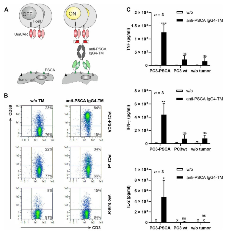
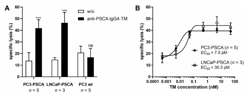
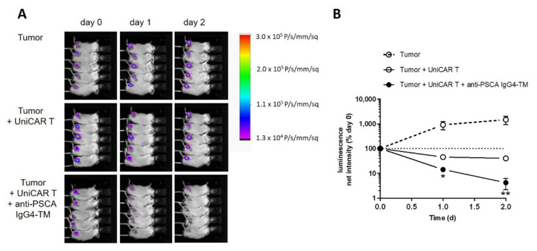
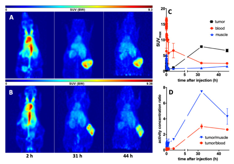
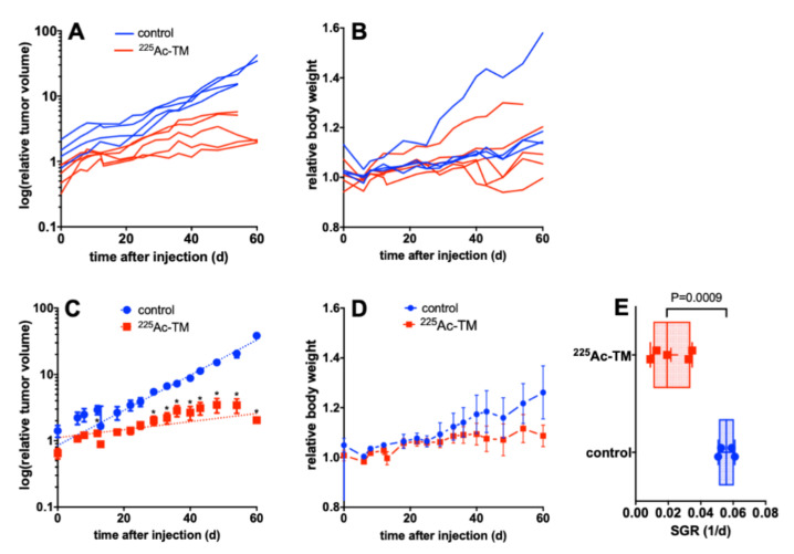
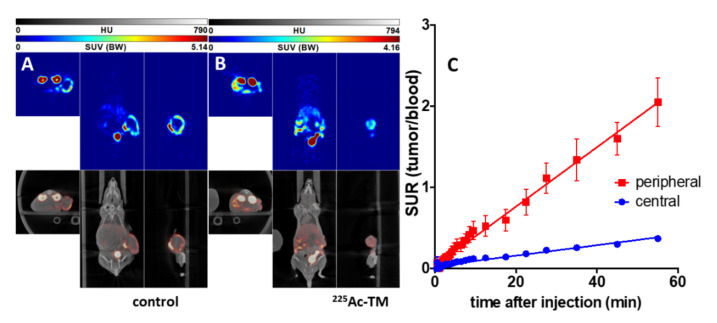
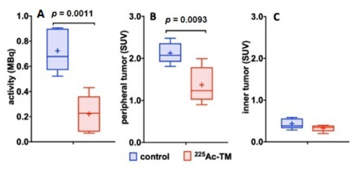
Due to its overexpression on the surface of prostate cancer (PCa) cells, the prostate stem cell antigen (PSCA) is a potential target for PCa diagnosis and therapy. Here we describe the development and functional characterization of a novel IgG4-based anti-PSCA antibody (Ab) derivative (anti-PSCA IgG4-TM) that is conjugated with the chelator DOTAGA. The anti-PSCA IgG4-TM represents a multimodal immunotheranostic compound that can be used (i) as a target module (TM) for UniCAR T cell-based immunotherapy, (ii) for diagnostic positron emission tomography (PET) imaging, and (iii) targeted alpha therapy. Cross-linkage of UniCAR T cells and PSCA-positive tumor cells via the anti-PSCA IgG4-TM results in efficient tumor cell lysis both in vitro and in vivo. After radiolabeling with 64Cu2+, the anti-PSCA IgG4-TM was successfully applied for high contrast PET imaging. In a PCa mouse model, it showed specific accumulation in PSCA-expressing tumors, while no uptake in other organs was observed. Additionally, the DOTAGA-conjugated anti-PSCA IgG4-TM was radiolabeled with 225Ac3+ and applied for targeted alpha therapy. A single injection of the 225Ac-labeled anti-PSCA IgG4-TM was able to significantly control tumor growth in experimental mice. Overall, the novel anti-PSCA IgG4-TM represents an attractive first member of a novel group of radio-/immunotheranostics that allows diagnostic imaging, endoradiotherapy, and CAR T cell immunotherapy.

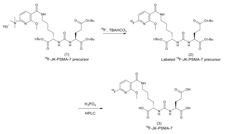
Keywords: (18)F-JK-PSMA-7; Ac-225; CAR T cell; Cu-64; IgG4; PSCA; PSMA; prostate cancer; theranostics.


Conflict of interest statement

The authors declare that there is no conflict of interest.

Figures

Figure 1
Figure 1
Functional characterization of VLc.5 and VLc.26 of the human anti-PSCA IgG1 Ab Ha1-4.121. (A) Schematic representation of the anti-PSCA scFvs Ha1-4.121 containing either the VLc.5 or VLc.26. C-terminally recombinant Abs were equipped with the E5B9-, myc- and hexahistidine-tag (His-tag). L, leader peptide; VL, variable domain of the light chain; VH, variable domain of the heavy chain; scFv, single-chain fragment variable. (B,C) Flow cytometry data of anti-PSCA scFv Ha1-4.121c.5 and anti-PSCA scFv Ha1-4.121c.26 using PC3-PSCA cells. After incubation of tumor cells with (B) 20 ng/µL or (C) increasing scFv concentrations, the binding of the respective scFv was detected via primary anti-La(5B9) mAb, and secondary goat anti-mouse IgG-PE Ab. (B) Histograms show percentage of stained cells (blue graphs) in comparison to the respective negative controls (black graphs). (C) For calculation of the apparent affinity of the scFvs towards PSCA, Ab concentrations were plotted against the relative mean fluorescence intensity (rel. MFI) values. Results of one representative experiment is shown; n.c., not calculable.
Figure 2
Figure 2
Biochemical characterization and binding properties of the recombinant anti-PSCA IgG4-TM. (A) The anti-PSCA scFv Ha1-4.1.2c.26 was fused to the hinge and Fc-region of human IgG4 molecules via flexible peptide linkers. In addition, the E5B9- and His-tag were fused to the C-terminus of the recombinant Ab. (B) The anti-PSCA IgG4-TM forms homodimers that are covalently connected via disulfide bounds in the hinge region. L, leader peptide; VL, variable domain of the light chain; VH, variable domain of the heavy chain; CH, constant domain of the heavy chain; scFv, single-chain fragment variable. (CE) After purification via protein A affinity chromatography, the elution fraction (E) containing the anti-PSCA IgG4-TM was analyzed via (C) SE-HPLC and (D,E) SDS-PAGE. (D) Proteins (TM monomer, Mw = 56 kDa) separated via SDS-PAGE were stained via Quick Coomassie® Stain solution to determine TM purity and concentration by means of a BSA standard. (E) After Western blotting, the anti-PSCA IgG4-TM was detected via its C-terminal His-tag. Uncropped WB in Figure S1. (F,G) Binding activity of the anti-PSCA IgG4-TM was evaluated by flow cytometry. PC3-PSCA cells were incubated with (F) 20 ng/µL or (G) increasing TM concentrations. Binding was detected via anti-His-PE Ab or primary anti-La(5B9) mAb, and secondary PE goat anti-mouse IgG (minimal x-reactivity) Ab. (F) Histograms show percentage of stained cells (blue graphs) in comparison to the respective negative controls (black graphs). (G) For calculation of affinity towards PSCA, Ab concentrations were plotted against the relative mean fluorescence intensity (rel. MFI) values. Results of three independent experiments are shown (rel. MFI ± SEM).
Figure 3
Figure 3
TM-mediated cross-linkage with tumor cells results in UniCAR T cell activation and cytokine release. (A) In the UniCAR T cell approach, T cells are engineered to express a universal chimeric antigen receptor (UniCAR) that recognizes the E5B9 peptide of the nuclear antigen La/SS-B. Thus, under physiological conditions UniCAR T cells are switched “OFF”. To engage the killing capabilities of UniCAR T cells against tumor cells (“ON”), an E5B9-tagged target module (TM) recognizing a tumor-associated antigen is required. (B,C) UniCAR T cells were incubated alone or with PC3-PSCA or PC3 wt cells at an E:T ratio of 5:1 in the presence or absence of 5 nM anti-PSCA IgG4-TM. After 24 h, (B) CD69 expression on UniCAR T cells and (C) secretion of TNF, IFN-γ and IL-2 were examined via flow cytometry or ELISA (x: not detectable), respectively. (B) Data of one representative experiment with one T cell donor are shown. (C) Summarized data of three different T cell donors are shown as mean ± SEM. (* p < 0.05, ** p < 0.01, *** p < 0.001 compared to samples w/o the anti-PSCA IgG4-TM; two-way ANOVA with post-hoc Šídák’s multiple comparisons test).
Figure 4
Figure 4
Redirection of UniCAR T cells for tumor cell killing via the novel anti-PSCA IgG4-TM. In 24 h standard chromium release assays, UniCAR T cells were incubated with PC3-PSCA, LNCaP-PSCA, or PC3 wt cells at an E:T ratio of 5:1 in the presence or absence of either (A) 5 nM or (B) decreasing concentrations of the anti-PSCA IgG4-TM. Mean specific lysis ± SEM of three or five different T cell donors are shown (*** p < 0.001 compared to samples w/o the anti-PSCA IgG4-TM; two-way ANOVA with post-hoc Šídák’s multiple comparisons test).
Figure 5
Figure 5
Evaluation of the immunotherapeutic potential of the anti-PSCA IgG4-TM in experimental mice. Immunodeficient NXG mice were subcutaneously injected with PC3-PSCA/PSMA Luc+ alone or in combination with UniCAR T cells in the absence or presence of the anti-PSCA IgG4-TM. (A) Luminescence images for each mouse after day 0, day 1, and day 2 are shown. (B) Quantitative analysis of luminescence signals. For each mouse, net intensities (P/s/mm2) were normalized to net intensities measured at day 0. Mean luminescence net intensities ± SEM for five mice per group are shown (* p < 0.05, ** p < 0.01, compared to the control group “Tumor + UniCAR T”, Student’s t-test).
Figure 6
Figure 6
Imaging and kinetics of the 64Cu-TM distribution in PC3-PSCA/PSMA Luc+ tumor bearing NMRI-Foxn1nu/nu mice. Maximum intensity projections of the PET studies of (A) mouse #1 and (B) mouse #2 at 2, 31, and 44 h after injection of the 64Cu-TM. (C) Activity–concentration–time curves in the tumor, blood, and muscle. (D) Tumor-to-muscle and tumor-to-blood ratios. Values are means ± SEM of two animals.
Figure 7
Figure 7
Targeted alpha therapy with the 225Ac-TM in a xenograft mouse model of PCa. (A,B) Individual or (C,D) averaged curves for (A,C) the tumor volume, and (B,D) body weight changes after DOTAGA-TM (control, n = 4) or 225Ac-TM (n = 5) injection in tumor-bearing mice. (E) The SGR of the relative tumor volumes were calculated with the exponential growth equation. SGR values for tumors of the control (0.0558 ± 0.0025, n = 4) and treatment group (0.0217 ± 0.0052, n = 5) correspond to doubling times of 12.4 and 32.0 days, respectively. Data are presented as means ± SEM. For each time point, the SGR of the single animals was analyzed with an unpaired t-test without assumption of consistent SD; the calculations were corrected for multiple comparisons using Holm-Sidak method; alpha of 0.05 was defined as ‘statistically significant’.
Figure 8
Figure 8
Scheme of the radiosynthesis of 18F-JK-PSMA-7.
Figure 9
Figure 9
18F-JK-PSMA-7 imaging of mice after targeted alpha therapy with the 225Ac-TM. Orthogonal sections of PET/CT studies of a representative (A) control and (B) 225Ac-TM-treated mouse with xenotransplanted PC3-PSCA/PSMA Luc+ tumors are shown. The PET studies were carried out at day 43 after treatment start and 2 h after single intravenous injection of 10 MBq 18F-JK-PSMA-7 with an imaging duration of 30 min. (C) Kinetics of the 18F-JK-PSMA-7 in the xenotransplanted untreated PC3-PSCA/PSMA Luc+ tumors (control) expressed as tumor uptake ratio (tumor-to-blood standard uptake ratio, SUR) (mean ± SEM of two animals of the control group) in the periphery (peripheral) and central part (central) of the tumors.
Figure 10
Figure 10
Quantitative comparison of the 18F-JK-PSMA-7 activity distribution. (A) 18F-JK-PSMA-7 activity amounts in the total tumor (MBq), (B) activity concentrations (SUV) in the periphery of the tumors, and (C) in the central part of the tumors are shown. The box and whiskers plots show mean SUV values for each group indicated as “+” in the control (n = 4) and 225Ac-TM treated group (n = 5).

Similar articles

Cited by

References

    1. Rebello R.J., Oing C., Knudsen K.E., Loeb S., Johnson D.C., Reiter R.E., Gillessen S., Van der Kwast T., Bristow R.G. Prostate cancer. Nat. Rev. Dis. Primers. 2021;7:9. doi: 10.1038/s41572-020-00243-0. - DOI - PubMed
    1. Farolfi A., Hadaschik B., Hamdy F.C., Herrmann K., Hofman M.S., Murphy D.G., Ost P., Padhani A.R., Fanti S. Positron Emission Tomography and Whole-body Magnetic Resonance Imaging for Metastasis-directed Therapy in Hormone-sensitive Oligometastatic Prostate Cancer After Primary Radical Treatment: A Systematic Review. Eur. Urol. Oncol. 2021;4:714–730. doi: 10.1016/j.euo.2021.02.003. - DOI - PubMed
    1. Debnath S., Zhou N., McLaughlin M., Rice S., Pillai A.K., Hao G., Sun X. PSMA-Targeting Imaging and Theranostic Agents-Current Status and Future Perspective. Int. J. Mol. Sci. 2022;23:1158. doi: 10.3390/ijms23031158. - DOI - PMC - PubMed
    1. Farolfi A., Mei R., Ali S., Castellucci P. Theragnostics in prostate cancer. Q. J. Nucl. Med. Mol. Imaging. 2021;65:333–341. doi: 10.23736/S1824-4785.21.03419-1. - DOI - PubMed
    1. FDA Approves First PSMA-Targeted PET Drug. [(accessed on 5 March 2022)];J. Nucl. Med. 2021 62:11N. Available online: https://pubmed.ncbi.nlm.nih.gov/33468545/ - PubMed

LinkOut - more resources