. 2022 Apr 14;14(8):1998.
doi: 10.3390/cancers14081998.

T-Cells Expressing a Highly Potent PRAME-Specific T-Cell Receptor in Combination with a Chimeric PD1-41BB Co-Stimulatory Receptor Show a Favorable Preclinical Safety Profile and Strong Anti-Tumor Reactivity

Affiliations

T-Cells Expressing a Highly Potent PRAME-Specific T-Cell Receptor in Combination with a Chimeric PD1-41BB Co-Stimulatory Receptor Show a Favorable Preclinical Safety Profile and Strong Anti-Tumor Reactivity

Nadja Sailer et al. Cancers (Basel). .

Abstract

The hostile tumor microenvironment (TME) is a major challenge for the treatment of solid tumors with T-cell receptor (TCR)-modified T-cells (TCR-Ts), as it negatively influences T-cell efficacy, fitness, and persistence. These negative influences are caused, among others, by the inhibitory checkpoint PD-1/PD-L1 axis. The Preferentially Expressed Antigen in Melanoma (PRAME) is a highly relevant cancer/testis antigen for TCR-T immunotherapy due to broad expression in multiple solid cancer indications. A TCR with high specificity and sensitivity for PRAME was isolated from non-tolerized T-cell repertoires and introduced into T-cells alongside a chimeric PD1-41BB receptor, consisting of the natural extracellular domain of PD-1 and the intracellular signaling domain of 4-1BB, turning an inhibitory pathway into a T-cell co-stimulatory pathway. The addition of PD1-41BB to CD8+ T-cells expressing the transgenic PRAME-TCR enhanced IFN-γ secretion, improved cytotoxic capacity, and prevented exhaustion upon repetitive re-challenge with tumor cells in vitro without altering the in vitro safety profile. Furthermore, a single dose of TCR-Ts co-expressing PD1-41BB was sufficient to clear a hard-to-treat melanoma xenograft in a mouse model, whereas TCR-Ts without PD1-41BB could not eradicate the PD-L1-positive tumors. This cutting-edge strategy supports development efforts to provide more effective TCR-T immunotherapies for the treatment of solid tumors.

Keywords: PD-1; PRAME; TCR-T cells; TME; cancer; immunotherapy.


Conflict of interest statement

M.S., D.B., C.G., K.M., and M.B. (Maja Bürdek) are employees and D.J.S. is a Managing Director of Medigene Immunotherapies GmbH, a subsidiary of Medigene AG, Planegg, Germany. N.S., I.F., C.K., M.B. (Monika Braun), S.W., and D.S. were employees of Medigene Immunotherapies GmbH during their contributions to this publication. N.S., M.B. (Monika Braun), and D.S. are current employees at Evotec; S.W. is a current employee at SCG Cell Therapy GmbH; I.F. is a current employee at Juno Therapeutics GmbH, a Bristol Myers Squibb Company; C.K. is a current employee at Intercept Pharma Deutschland GmbH.

Figures

Figure 1
Figure 1
Isolation and selection of a lead PRAME-SLL-TCR from a non-tolerized T-cell repertoire. (A) CD8+ T-cells from HLA-A*02-negative healthy donors were stimulated using autologous monocyte-derived mature dendritic cells (mDCs) transfected with ivtRNA encoding PRAME and HLA-A*02:01. Dot blot shows staining with PE (x-axis) and APC (y-axis) labeled PRAME-multimers used for single-cell FACS. Percentage of double-positive cells is indicated. Promising TCRs were reconstructed and transferred into effector CD8+ recipient T-cells by retroviral transduction. Based on multi-parameter screening (triage process), four promising TCR candidates (TCR-027-004, TCR-027-085, TCR-038-038, and TCR-061-119) were selected from more than 30 analyzed specific TCRs. (B) Functional avidity of TCR-transgenic T-cells was analyzed in co-cultures with peptide-loaded T2 cells. Cells were cultured at an E:T ratio of 1:1. The graph shows nonlinear regression curves of relative IFN-γ values. Half maximal IFN-γ secretion is indicated by the dashed line at 50%. The data represent the means of duplicates measured at each peptide concentration. (C) IFN-γ concentrations released by the different TCR-Ts after co-culture with HLA-A*02-positive tumor cell lines that were either PRAME-positive (SKMel23, Mel624.38 and MelA375) or PRAME-negative (647-V, MCF-7 and Colo678). Untransduced (UT) T-cells served as negative control. IFN-γ values are shown as the mean of duplicates. Cells were cultured at an E:T ratio of 4:1 except for 647-V cells, which were cultured at an E:T ratio of 8:1. (D) Cytotoxicity of TCR-Ts expressing different PRAME-TCRs against fluorescently labeled HLA-A*02-positive tumor cell lines (PRAME-negative: 647-V; PRAME-positive: SKMel23, MelA375), assessed using a live-cell imaging system (IncuCyte® ZOOM, Sartorius). Loss of red fluorescence visualized tumor cell apoptosis. UT effectors served as negative control. Cells were cultured at an E:T ratio of 4:1 except for 647-V cells, which were cultured at an E:T ratio of 8:1. Mean of cell counts using triplicates are shown over time. Statistical significance was calculated with one-way ANOVA with multiple comparisons (* p < 0.03, ** p < 0.002, **** p < 0.0001). Experiments in (BD) were repeated three times with T-cells derived from different donors; data from one representative donor are shown. (E,F) 5 × 10⁶ Mel624.38 or MelA375_PD-L1 cells were injected s.c. into immunodeficient (NOD/Shi-scid/IL-2Rγnull) mice. Mice were treated on day 14 for Mel624.38 and day 7 for MelA375_PD-L1, respectively, with T-cells expressing TCR-027-004 or UT T-cells (5–6 mice per group) and (E) tumor growth and (F) survival of mice was followed until day 67 after tumor cell injection. Statistical significance between UT and TCR was calculated using the log-rank Mantel–Cox test (Mel624.38 p = 0.0018 and MelA375_PD-L1 p = 0.3597).
Figure 2
Figure 2
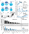
Co-expressing PD1-41BB on PRAME-TCR-Ts increases effector function in response to PRAME-positive tumor cell lines. (A) Interaction of wildtype PD1 expressed by T-cells with PD-L1 on tumor cells results in inhibition of T-cell activity and can lead to exhaustion and apoptosis. In contrast, the presence of the chimeric PD1-41BB co-stimulatory receptor on TCR-Ts can turn this inhibition into activation leading to improved effector functions, survival, and longevity. (B) Flow cytometric analysis of TCR-Ts with or without PD1-41BB for expression of the PRAME-TCR (TRBV09) (PE) and PD1-41BB (PD1) (Alexa Fluor-647). The analysis was repeated for transduced T-cells derived from 4 different donors. Data from one representative donor are shown. (C) Upper part: PRAME-RNA expression levels of tumor cell lines derived from different indications were analyzed by qPCR. Tumor cell lines are ordered from high to low expression from left to the right. Bars represent means of triplicates ± mean deviation. PD-L1 expression levels with and without IFN-γ treatment were analyzed by flow cytometry. PD-L1 status is indicated below the graph: (TD) transduced, (ind) IFN-γ inducible, (end) endogenously expressed. Lower part: TCR-Ts with or without PD1-41BB were co-cultured with the corresponding tumor cell lines at an E:T ratio of 1:1 and IFN-γ levels were determined 24 h after co-culture by ELISA. IFN-γ values are shown as the mean of triplicates ± mean deviation. (D) Cytotoxicity of TCR-Ts with or without PD1-41BB against red fluorescently labeled 3D tumor cell spheroids was monitored over 16 days using the IncuCyte S3® device. T-cells were re-challenged with fresh spheroids on days 3, 7, 10 and 13, indicated by black arrows. PD-L1-transduced SKMel23 (PRAME-positive), endogenously PD-L1-expressing NCI-H1650 (PRAME-positive), and endogenously PD-L1-expressing 647-V (PRAME-negative) were used as target cells. UT T-cells served as control. Limit of analysis for UT T-cells was reached when red fluorescence could no longer be reliably calculated due to an excess of tumor cells in the well (#). Statistical significance was calculated from end-point values with unpaired t-test (TCR vs. TCR-PD1-41BB; p = 0.0076 (**) for SKMel23_PD-L1; p = 0.0081 (**) for NCH-H1650). Experiments in (BD) were repeated four times with T-cells derived from different donors; data with cells from one representative donor are shown.
Figure 3
Figure 3
Co-expression of PD1-41BB does not change the in vitro safety profile of the lead PRAME-SLL-TCR. (A) Functional avidity of CD8+ TCR-Ts, with or without PD1-41BB, following stimulation with PD-L1-transduced T2 cells loaded with graded amounts of PRAME peptide (10−10 M–10−5 M). Cells were cultured at an E:T ratio of 1:1. The graph shows nonlinear regression curves of relative IFN-γ release. Peptide concentration needed for half maximal IFN-γ secretion is indicated by the dashed line. The data represent means of duplicates measured at each peptide concentration. (B) Left part: The recognition of 191 peptides with high sequence homology with the specific PRAME-SLL peptide by CD8+ TCR-Ts with or without PD1-41BB was analyzed. Peptides were loaded onto T2_PD-L1 cells at a concentration of 10−6 M. IFN-γ release data for 30 peptides are shown as an example; data for all peptides are shown in Figure S2. Right part: PRAME-negative, PD-L1-expressing SNB-19 cells were transfected with RNA constructs encoding either a midigene (~400 bp) coding for single peptides (SLL, 1, 26, 66) or minigene (~90 bp per peptide) constructs (MG) coding for up to five variant peptides and co-cultured with effector cells. All constructs included an epitope recognized by a positive control TCR. UT T-cells served as negative control. For read-out, supernatants were harvested after 20 h and analyzed by IFN-γ ELISA. The data represent means of duplicates. (C) IFN-γ released by CD8+ TCR-Ts with or without PD1-41BB co-cultured with 36 LCLs covering frequent HLA-A, -B, and -C alleles. UT CD8+ T-cells served as negative control, and PRAME-SLL-peptide-loaded HLA-A*02:01 positive LCLs were used as positive control. (D) IFN-γ released by CD8+ TCR-Ts with or without PD1-41BB co-cultured with normal cells derived from critical healthy tissues. As positive controls, cells were loaded with PRAME SLL-peptide. HREpC: human renal epithelial cells, HRCEpC: human renal cortical epithelial cells, RPTEC: renal proximal tubule epithelial cells, NHBE: normal human bronchial epithelial cells, NHLF: normal human lung fibroblasts, HOB: human osteoblasts, Mono: monocytes, iDC: immature dendritic cells, mDC: mature dendritic cells, iCardio: iCell Cardiomyocytes. The data represent means of duplicates. (E) Cytotoxicity of CD8+ TCR-Ts with or without PD1-41BB against PRAME-positive PD-L1-negative (red) and PRAME-negative PD-L1-positive (green) 3D tumor cell spheroids. Fresh PRAME-positive Mel624.38 spheroids (red) were added to the co-cultures on days 3 and 6, indicated by black arrows. For days 3 and 6 pictures, before and after adding fresh tumor cell spheroids are shown. Experiments in A–E were repeated at least three times with T-cells derived from different donors; data with cells from one representative donor are shown.
Figure 4
Figure 4
TCR-Ts co-expressing PD1-41BB reject tumor cells in vivo and show a superior poly-cytokine profile. (A) 5 × 10⁶ MelA375_PD-L1 cells were injected s.c. into immunodeficient (NOD/Shi-scid/IL-2Rγnull) mice. Mice were treated on day seven either with 10 × 106 PRAME-TCR-positive CD8+ TCR-Ts with (7 mice) or without (7 mice) PD1-41BB (14 × 106 cells in total per mouse) or 14 × 106 UT T-cells (6 mice), and tumor growth (left panel) and survival (right panel) was followed for 60 days. Statistical significance between TCR and TCR_PD1-41BB was calculated using the log-rank Mantel–Cox test (p = 0.0003, ***). (B) Co-cultures of MelA375_PD-L1 cells and TCR-Ts, with and without PD1-41BB, were performed in poly-L-lysine pre-coated 24 well plates. After 20 h, T-cells were harvested, enriched for CD8 expression by MACS, and applied onto IsoCode Chips. Polyfunctional strength index (PSI), which combines the analysis of identity and quantity of secreted cytokines/proteins, is depicted. Analysis is based on all cytokines/proteins which were detected by the Single-Cell PF Strength Panel kit designed for 32 different proteins. Experiment was repeated two times with T-cells derived from different donors. Data from one representative donor are shown.

References

    1. Jiang Y., Li Y., Zhu B. T-cell exhaustion in the tumor microenvironment. Cell Death Dis. 2015;6:e1792. doi: 10.1038/cddis.2015.162. - DOI - PMC - PubMed
    1. Maimela N.R., Liu S., Zhang Y. Fates of CD8+ T cells in Tumor Microenvironment. Comput. Struct. Biotechnol. J. 2018;17:1–13. doi: 10.1016/j.csbj.2018.11.004. - DOI - PMC - PubMed
    1. Topalian S.L., Hodi F.S., Brahmer J.R., Gettinger S.N., Smith D.C., McDermott D.F., Powderly J.D., Carvajal R.D., Sosman J.A., Atkins M.B., et al. Safety, Activity, and Immune Correlates of Anti-PD-1 Antibody in Cancer. N. Engl. J. Med. 2012;366:2443–2454. doi: 10.1056/NEJMoa1200690. - DOI - PMC - PubMed
    1. Sharma P., Allison J.P. Immune Checkpoint Targeting in Cancer Therapy: Toward Combination Strategies with Curative Potential. Cell. 2015;161:205–214. doi: 10.1016/j.cell.2015.03.030. - DOI - PMC - PubMed
    1. Callahan M.K., Postow M.A., Wolchok J.D. CTLA-4 and PD-1 Pathway Blockade: Combinations in the Clinic. Front. Oncol. 2015;4:385. doi: 10.3389/fonc.2014.00385. - DOI - PMC - PubMed