Review
. 2022 Apr 15;14(8):2004.
doi: 10.3390/cancers14082004.

Deep-Tissue Activation of Photonanomedicines: An Update and Clinical Perspectives

Affiliations
Review

Deep-Tissue Activation of Photonanomedicines: An Update and Clinical Perspectives

Nimit Shah et al. Cancers (Basel). .

Abstract

With the continued development of nanomaterials over the past two decades, specialized photonanomedicines (light-activable nanomedicines, PNMs) have evolved to become excitable by alternative energy sources that typically penetrate tissue deeper than visible light. These sources include electromagnetic radiation lying outside the visible near-infrared spectrum, high energy particles, and acoustic waves, amongst others. Various direct activation mechanisms have leveraged unique facets of specialized nanomaterials, such as upconversion, scintillation, and radiosensitization, as well as several others, in order to activate PNMs. Other indirect activation mechanisms have leveraged the effect of the interaction of deeply penetrating energy sources with tissue in order to activate proximal PNMs. These indirect mechanisms include sonoluminescence and Cerenkov radiation. Such direct and indirect deep-tissue activation has been explored extensively in the preclinical setting to facilitate deep-tissue anticancer photodynamic therapy (PDT); however, clinical translation of these approaches is yet to be explored. This review provides a summary of the state of the art in deep-tissue excitation of PNMs and explores the translatability of such excitation mechanisms towards their clinical adoption. A special emphasis is placed on how current clinical instrumentation can be repurposed to achieve deep-tissue PDT with the mechanisms discussed in this review, thereby further expediting the translation of these highly promising strategies.

Keywords: Cerenkov radiation; X-ray; bioluminescence; chemiluminescence; photodynamic therapy; photonanomedicines; proton therapy; sonodynamic therapy; tumor; two-photon; ultrasound therapy; upconversion.


Conflict of interest statement

The authors declare no conflict of interest.

Figures
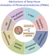
Figure 1
Figure 1
A summary of various mechanisms of deep-tissue activation of photonanomedicines (PNMs) using alternative tissue permeating energy sources that will be discussed in this review. Deep-tissue activation of the PNM leads to phototoxicity or photodynamic priming of the tumor microenvironment that can enable and potentiate combinatorial modalities. (Figure is created from Biorender.com using an Academic License).
Figure 2
Figure 2
Schematic representation of two-photon-mediated photodynamic therapy (PDT). (A) A Jablonski diagram of two-photon excitation which shows how PNM can be activated using two-photon excitation (TPE). (B) The administration of PNMs into the animal model which accumulate at the tumor site. When activated by TPE, they generate reactive molecular species (RMS) which cause biomodulation and ultimately cell death. (Figure is created from Biorender.com using an Academic License.)
Figure 3
Figure 3
Schematic representation of general mechanism of upconversion PDT. (A) A Jablonski diagram demonstrating the upconversion process whereby two consecutive long wavelength excitations (hv1 and hv2) of upconverting nanoparticles (UCNPs) result in short wavelength emission (hv3) from higher energy levels. (B) Administration and circulation of UCNP-based PNMs within the blood vessels and their uptake in cancer cells. Furthermore, the general mechanism of cell killing through production of reactive molecular species is demonstrated. UCNPs are activated by NIR light which further emits visible light and excites PSs for upconversion PDT. (Figure is created from Biorender.com using an Academic License.)
Figure 4
Figure 4
(A) Synthesis stages of UCNPs-ZnPc constructs prepared by Xia et al. (B) Cell viability of HeLa cells treated with and without irradiation (0.39 W/cm2 at 980 nm) along with different concentration of UCNPs-ZnPc/FA. (C) Hepa1-6 tumor volumes in different groups of treatment including upconversion PDT. (D) Images of tumor and mice in the same treatment groups as in (C). Reprinted and adapted with permission from [27]. Copyright (2014) American Chemical Society.
Figure 5
Figure 5
Schematic representation of the general mechanism of action for direct X-PDT without the use of a nanoscintillator intermediate. (Figure is created from Biorender.com using an Academic License.)
Figure 6
Figure 6
A schematic diagram that shows the general mechanism of nanoscintillator facilitated X-PDT. The figure represents the administration of nanoscintillator-based PNM into the animal model and its uptake in tumor tissue. Furthermore, it demonstrates the general X-PDT process using nanoscintillator systems as fluorescence resonance energy transfer (FRET) donors for the generation of reactive molecular species (RMS). (Figure is created from Biorender.com using an Academic License.)
Figure 7
Figure 7
(A) Schematic of X-PDT using a SrAl2O4:Eu2+ nanoscintillator with the PS MC540. (B) Cell viabilities of U87MG (human glioblastoma) with treatments at different groups with and without X-rays and nanoscintillators (n = 4). (C) U87MG tumor growth curves following X-PDT (V/V0%, n = 5). (D) Changes in animal body weight following X-PDT. (E) H&E staining of tumor tissues following X-PDT. Reprinted and adapted with permission from [36]. Copyright (2015) American Chemical Society.
Figure 8
Figure 8
Schematic representation of Cerenkov-radiation-induced PDT represents the injection of a PNM system into the animal model and its accumulation in tumors. Furthermore, it represents the process of generating reactive molecular species through activation of a PS by Cerenkov radiation, ultimately leading to cell death. (Figure is created from Biorender.com using an Academic License).
Figure 9
Figure 9
(A) A representation of the stepwise synthesis of the [89Zr]HMSN-Ce6 PNM complex for Cerenkov-radiation-induced PDT. (B) Cell viability of 4T1 cells when treated with 89Zr, HMSNs, HMSNs-Ce6, and [89Zr] HMSN-Ce6. (C) Tumor (mammary carcinoma) growth curves of different treatment groups: control group (black), [89Zr] HMSN (blue), HMSN-Ce6 (green), and [89Zr]HMSN-Ce6 (red), with n = 4. Error bars are SD. Statistical analysis was calculated by the student’s t test (***, p < 0.001; **, p < 0.01) [60]. Copyright (2016) American Chemical Society.
Figure 10
Figure 10
Schematic representation of chemiluminescence resonance energy transfer (CRET) and bioluminescence resonance energy transfer (BRET)-mediated PDT. (Figure is created from Biorender.com using an Academic License).
Figure 11
Figure 11
(A) A schematic representing the synthesis of PLGA-RB nanoparticles and mechanism of ROS generation following BRET. (B) Data from in vivo animal studies of the PLGA-RB nanoparticles by BRET-PDT. (B(a)) Tumor (mouse hepatocellular carcinoma) volume of 5 groups. Data are shown as the mean ± SD. (B(b)) Tumor volumes from 0 and 14 d. (c) Body weight of animals in all 5 groups. (d) Images of tumors excised on 24 d after treatment. (PLGA RB nanoparticles (PLGA-RB NPs): 40 μg/mL, PLGA-RB NPs conjugated with 20 μg/mL luciferase (NP-luciferase), luciferin: 60 μmol/L.) Reprinted and adapted with permission from [74]. Copyright (2018) American Chemical Society.
Figure 12
Figure 12
Schematic representation demonstrating the processes behind sonodynamic therapy (SDT) using PNMs. (Figure is created from Biorender.com using an Academic License).

Similar articles

Cited by

References

    1. Bhandari C., Guirguis M., Savan N.A., Shrivastava N., Oliveira S., Hasan T., Obaid G. What NIR Photodynamic Activation Offers Molecular Targeted Nanomedicines: Perspectives into the Conundrum of Tumor Specificity and Selectivity. Nano Today. 2021;36:101052. doi: 10.1016/j.nantod.2020.101052. - DOI - PMC - PubMed
    1. Hu J.J., Lei Q., Zhang X.Z. Recent Advances in Photonanomedicines for Enhanced Cancer Photodynamic Therapy. Prog. Mater. Sci. 2020;114:100685. doi: 10.1016/j.pmatsci.2020.100685. - DOI
    1. Obaid G., Broekgaarden M., Bulin A.-L., Huang H.-C., Kuriakose J., Liu J., Hasan T. Photonanomedicine: A Convergence of Photodynamic Therapy and Nanotechnology. Nanoscale. 2016;8:12471–12503. doi: 10.1039/C5NR08691D. - DOI - PMC - PubMed
    1. Obaid G., Jin W., Bano S., Kessel D., Hasan T. Nanolipid Formulations of Benzoporphyrin Derivative: Exploring the Dependence of Nanoconstruct Photophysics and Photochemistry on Their Therapeutic Index in Ovarian Cancer Cells. Photochem. Photobiol. 2019;95:364–377. doi: 10.1111/php.13002. - DOI - PMC - PubMed
    1. Spring B.Q., Sears R.B., Zheng L.Z., Mai Z., Watanabe R., Sherwood M.E., Schoenfeld D.A., Pogue B.W., Pereira S.P., Villa E. A Photoactivable Multi-Inhibitor Nanoliposome for Tumour Control and Simultaneous Inhibition of Treatment Escape Pathways. Nat. Nanotechnol. 2016;11:378–387. doi: 10.1038/nnano.2015.311. - DOI - PMC - PubMed

LinkOut - more resources