

ERα36-High Cancer-Associated Fibroblasts as an Unfavorable Factor in Triple-Negative Breast Cancer
- PMID: 35454913
- PMCID: PMC9024776
- DOI: 10.3390/cancers14082005
ERα36-High Cancer-Associated Fibroblasts as an Unfavorable Factor in Triple-Negative Breast Cancer
Abstract
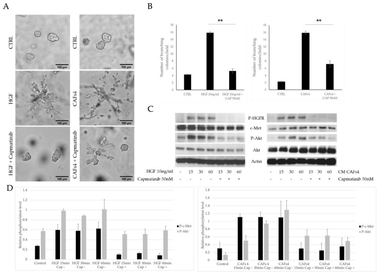
Background: Cancer-associated fibroblasts (CAFs) are the most abundant cell type in the tumor microenvironment (TME). Estrogen receptor alpha 36 (ERα36), the alternatively spliced variant of ERα, is described as an unfavorable factor when expressed in cancer cells. ERα can be expressed also in CAFs; however, the role of ERα36 in CAFs is unknown. Methods: Four CAF cultures were isolated from chemotherapy-naïve BC patients and characterized for ERα36 expression and the NanoString gene expression panel using isolated RNA. Conditioned media from CAF cultures were used to assess the influence of CAFs on triple-negative breast cancer (TNBC) cells using a matrigel 3D culture assay. Results: We found that ERα36high CAFs significantly induced the branching of TNBC cells in vitro (p < 0.001). They also produced a set of pro-tumorigenic cytokines compared to ERα36low CAFs, among which hepatocyte growth factor (HGF) was the main inducer of TNBC cell invasive phenotype in vitro (p < 0.001). Tumor stroma rich in ERα36high CAFs was correlated with high Ki67 expression (p = 0.041) and tumor-associated macrophages markers (CD68 and CD163, p = 0.041 for both). HGF was found to be an unfavorable prognostic factor in TCGA database analysis (p = 0.03 for DFS and p = 0.04 for OS). Conclusions: Breast cancer-associated fibroblasts represent distinct subtypes based on ERα36 expression. We propose that ERα36high CAFs could account for an unfavorable prognosis for TNBC patients.
Keywords: breast cancer; cancer-associated fibroblasts; estrogen receptor alpha 36; triple-negative breast cancer.
Conflict of interest statement
The authors declare no conflict of interest.
Figures


Similar articles
-
The role of CAFs in therapeutic resistance in triple negative breast cancer: an emerging challenge.Front Mol Biosci. 2025 Mar 31;12:1568865. doi: 10.3389/fmolb.2025.1568865. eCollection 2025. Front Mol Biosci. 2025. PMID: 40230452 Free PMC article. Review.
-
Alpha-smooth muscle actin-positive cancer-associated fibroblasts secreting osteopontin promote growth of luminal breast cancer.Cell Mol Biol Lett. 2022 Jun 11;27(1):45. doi: 10.1186/s11658-022-00351-7. Cell Mol Biol Lett. 2022. PMID: 35690734 Free PMC article.
-
Effects of cytokines derived from cancer-associated fibroblasts on androgen synthetic enzymes in estrogen receptor-negative breast carcinoma.Breast Cancer Res Treat. 2017 Dec;166(3):709-723. doi: 10.1007/s10549-017-4464-5. Epub 2017 Aug 22. Breast Cancer Res Treat. 2017. PMID: 28831645
-
Landscape of cancer-associated fibroblasts identifies the secreted biglycan as a protumor and immunosuppressive factor in triple-negative breast cancer.Oncoimmunology. 2022 Jan 3;11(1):2020984. doi: 10.1080/2162402X.2021.2020984. eCollection 2022. Oncoimmunology. 2022. PMID: 35003899 Free PMC article.
-
CAF-Associated Genes in Breast Cancer for Novel Therapeutic Strategies.Biomedicines. 2024 Aug 29;12(9):1964. doi: 10.3390/biomedicines12091964. Biomedicines. 2024. PMID: 39335478 Free PMC article. Review.
Cited by
-
Integrated analysis of fibroblasts molecular features in papillary thyroid cancer combining single-cell and bulk RNA sequencing technology.Front Endocrinol (Lausanne). 2022 Oct 26;13:1019072. doi: 10.3389/fendo.2022.1019072. eCollection 2022. Front Endocrinol (Lausanne). 2022. PMID: 36387901 Free PMC article.
-
Regulation of Stromal Cells by Sex Steroid Hormones in the Breast Cancer Microenvironment.Cancers (Basel). 2024 Dec 2;16(23):4043. doi: 10.3390/cancers16234043. Cancers (Basel). 2024. PMID: 39682229 Free PMC article. Review.
-
The role of CAFs in therapeutic resistance in triple negative breast cancer: an emerging challenge.Front Mol Biosci. 2025 Mar 31;12:1568865. doi: 10.3389/fmolb.2025.1568865. eCollection 2025. Front Mol Biosci. 2025. PMID: 40230452 Free PMC article. Review.
-
The untapped potential of radiation and immunotherapy for hormone receptor-positive breast cancer.NPJ Breast Cancer. 2025 Jul 24;11(1):77. doi: 10.1038/s41523-025-00796-x. NPJ Breast Cancer. 2025. PMID: 40707495 Free PMC article. Review.
References
-
- Li X., Yang J., Peng L., Sahin A.A., Huo L., Ward K.C., O’Regan R., Torres M.A., Meisel J.L. Triple-negative breast cancer has worse overall survival and cause-specific survival than non-triple-negative breast cancer. Breast Cancer Res. Treat. 2017;161:279–287. doi: 10.1007/s10549-016-4059-6. - DOI - PubMed
-
- Goldhirsch A., Wood W.C., Coates A.S., Gelber R.D., Thurlimann B., Senn H.J. Strategies for subtypes—Dealing with the diversity of breast cancer: Highlights of the St. Gallen International Expert Consensus on the primary therapy of early breast cancer 2011. Ann. Oncol. 2011;22:1736–1747. doi: 10.1093/annonc/mdr304. - DOI - PMC - PubMed
Grants and funding
LinkOut - more resources
Full Text Sources
Molecular Biology Databases
Research Materials
