

Capillaroscopy and Immunological Profile in Systemic Sclerosis
- PMID: 35454989
- PMCID: PMC9024594
- DOI: 10.3390/life12040498
Capillaroscopy and Immunological Profile in Systemic Sclerosis
Abstract
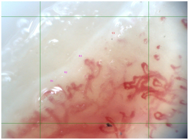
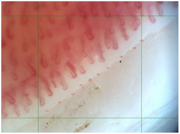
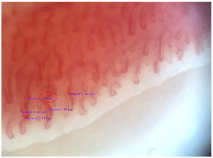
Introduction: Data on the associations between capillaroscopic changes and diagnostic systemic-sclerosis (SSc)-related antibodies are scarce. Presence of such correlation would improve current knowledge about the disease’s pathogenesis by revealing the mechanisms of microangiopathy. The microvascular pathology of SSc is a hallmark of the disease, and immunological abnormalities probably contribute to its development. Patients and methods: 19 patients with definite diagnosis of SSc were included in the current pilot study; 16 had limited and 3 had diffuse cutaneous involvement; their mean age was 51.56 ± 15.07 years. All patients exhibited symptoms of Raynaud’s phenomenon of the fingers. A “scleroderma” type capillaroscopic pattern was classified according to the staging suggested by Cutolo et al. (2000): “early”, “active” or ”late” phase. In the presence of different degrees of capillaroscopic changes in different fingers, the most-advanced microvascular pathology was chosen for classification. In cases without capillaroscopic features of microangiopathy, the findings were categorized as normal or nonspecific (dilated, tortuous capillaries, and/or hemorrhages). Indirect immunofluorescence on HEp-2 cells was performed as the gold-standard screening method for the detection of antinuclear autoantibodies (ANA), and determination of the immunofluorescent staining pattern (anti-cell pattern) was in accordance with the International Consensus on ANA Patterns. Scleroderma-associated autoantibodies in the patients’ serum were assessed using line immunoblot assay for detection of autoantibodies to 13 scleroderma-associated autoantigens: Scl-70, CENP A, CENP B, RP11/RNAP-III, RP155/RNAP-III, fibrillarin, NOR-90, Th/To, PM-Scl100, PM-Scl75, Ku, PDGFR, and Ro-52. Results: In 73.7% (n = 14) of the examined patients, “scleroderma” type capillaroscopic changes were found, and in 26.3% (n = 5), capillaroscopic features of microangiopathy were absent (nonspecific changes, n = 3; normal findings, n = 2). In SSc patients with positive anti-Scl-70 (n = 7) antibodies, significantly lower mean capillary density was observed along with a higher frequency of “active” and “late” phase capillaroscopic changes as compared to the anti-Scl-70-negative patients (p < 0.05). Anti-RNAP III−155 positive patients (n = 4) had significantly higher mean capillary density than anti-RNAP III−155 negative patients (n = 15). In three of the anti-RNAP III−155-positive cases, capillaroscopic features of microangiopathy were not detected, and in one case there was an “early” phase “scleroderma” pattern. Conclusion: In the current pilot study, the association between more advanced capillaroscopic changes and the presence of anti-Scl-70 autoantibodies was confirmed. As a novel observation, positive anti-RNAP III−155 antibodies were found in SSc patients with or without early microangiopathy. The question of associations between microvascular changes in SSc and other SSc-related autoantibodies requires further research.
Keywords: autoantibodies; capillaroscopy; systemic sclerosis.
Conflict of interest statement
The authors declare no conflict of interest.
Figures
References
-
- Van Den Hoogen F., Khanna D., Fransen J., Johnson S.R., Baron M., Tyndall A., Matucci-Cerinic M., Naden R.P., Medsger T.A., Jr., Carreira P.E., et al. 2013 classification criteria for systemic sclerosis: An American College of Rheumatology/European League Against Rheumatism collaborative initiative. Ann. Rheum. Dis. 2013;71:1747–1755. doi: 10.1136/annrheumdis-2013-204424. - DOI - PubMed
-
- Van Den Hoogen F., Khanna D., Fransen J., Johnson S.R., Baron M., Tyndall A., Matucci-Cerinic M., Naden R.P., Medsger T.A., Jr., Carreira P.E., et al. 2013 classification criteria for systemic sclerosis: An American College of Rheumatology/European League Against Rheumatism collaborative initiative. Arthritis Rheum. 2013;65:2737–2747. doi: 10.1002/art.38098. - DOI - PMC - PubMed
-
- Maricq H.R., Harper F.E., Khan M.M., Tan E.M., LeRoy E.C. Microvascular abnormalities as possible predictors of disease subsets in Raynaud phenomenon and early connective tissue disease. Clin. Exp. Rheumatol. 1983;1:195–205. - PubMed
LinkOut - more resources
Full Text Sources
Research Materials
Miscellaneous
