. 2022 Apr 4;12(4):531.
doi: 10.3390/life12040531.

Effects of Electrical Stimulation on the Signal Transduction-Related Proteins, c-Src and Focal Adhesion Kinase, in Fibroblasts

Affiliations

Effects of Electrical Stimulation on the Signal Transduction-Related Proteins, c-Src and Focal Adhesion Kinase, in Fibroblasts

Kazuo Katoh. Life (Basel). .

Abstract

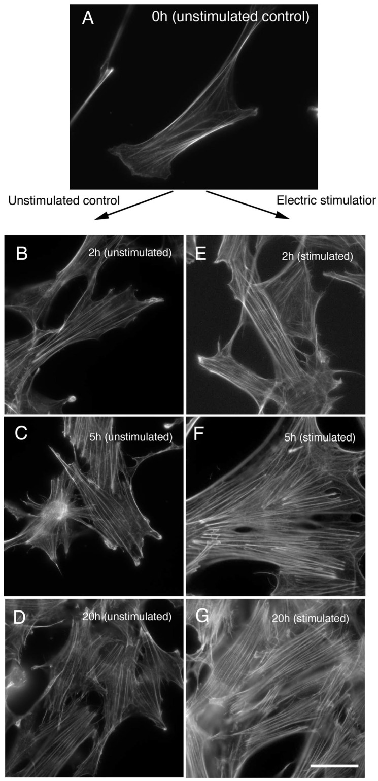
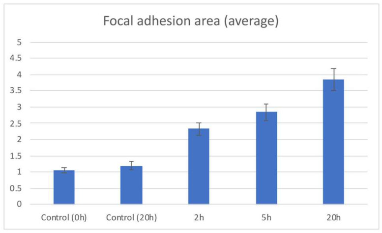
Electrical stimulation of the skin and muscles, e.g., in the fields of rehabilitation medicine and acupuncture, is known to locally increase blood flow and metabolism, and thus have beneficial health effects. However, little is known about the changes in cellular morphology or regulation of the localization of specific proteins in response to electrical stimuli. The present study was performed to examine the effects of electrical stimulation on the cytoskeletal system of cultured fibroblasts. Following application of electrical stimulation to cultured fibroblastic cells for a period of about 2 h, the stress fibers in the cells became thicker and the cells showed a contracted appearance. Cells were subjected to periodic electrical stimulation for 0 (unstimulated control), 2, 5, or 20 h. The stress fibers showed an increase in thickness within 2 h, and became gradually thicker until 20 h. In addition, the focal adhesions and stress fibers were enlarged after 2 h of continuous stimulation, and both stress fibers and focal adhesions became larger and thicker after 20 h of periodic stimulation. Cells showed increased staining of focal adhesions with anti-phosphotyrosine antibody (PY-20) after electrical stimulation. Cells also showed increased staining of tyrosine-phosphorylated focal adhesion kinase (FAK) (pY397) and tyrosine-phosphorylated c-Src (pY418), indicating that electrical stimulation affected signal transduction-related proteins.

Keywords: c-Src; cytoskeleton; electrical stimulation; focal adhesion kinase.


Conflict of interest statement

The author declares no conflict of interest.

Figures

Figure 1
Figure 1
Apparatus for providing single-pulse electrical stimulation to cells and simple stimulation device. We developed a device using an electronic circuit and platinum wire to apply electrical stimulation to cultured cells, and confirmed that it could apply electrical stimulation to the cells (A). A schematic illustration is shown in (B).
Figure 2
Figure 2
Changes in stress fibers during electrical stimulation of cells. The stress fibers of fibroblasts were significantly altered with the application of periodic electrical stimuli at 50 V, 60 times/min. Electrical stimuli were applied to the cells for 0 (control) (A), 2 (E), 5 (F), or 20 h (overnight) (G). The stress fibers began to increase in number after about 1 h of electrical stimulation. The stress fibers became remarkably thicker after about 2 h of electrical stimulation (compare (B) unstimulated control, and (E) electric stimulation for 2 h). The stress fibers became more thicker after about 5 h of electrical stimulation (compare (C) unstimulated control, and (F) electric stimulation for 5 h). After 20 h of periodic electrical stimulation, the number of stress fibers remained the same, but the fibers increased in thickness (compare (D) unstimulated control for 20 h, and (G) electric stimulation for 20 h). Bars: 20 μm.
Figure 3
Figure 3
Changes in focal adhesions after electrical stimulation of cells. Fibroblasts were stained with monoclonal anti-paxillin antibody, and the focal adhesions were examined after periodic electrical stimulation (0, 2, 5, 20 h). Focal adhesions are gradually enlarged in size, according to the electrical stimulation for 2 (E), 5 (F), and 20 (G) hours. (A) 0 h (unstimulated control); (B) 2 h (unstimulated control); (C) 5 h (unstimulated control); (D) 20 h (unstimulated control); (E) 2 h (electrical stimulation); (F) 5 h (electrical stimulation); (G) 20 h (electrical stimulation). Bars: 20 μm.
Figure 4
Figure 4
Increase in focal adhesions and accumulation of phosphotyrosine-containing proteins by electrical stimulation. After 2 h of electrical stimulation of GFP-paxillin-transfected cells, the focal adhesions began to enlarge (DF). After 5 (GI) to 20 h (JL) of periodic electrical stimulation, the focal adhesions showed intense staining with monoclonal anti-phosphotyrosine antibody (PY-20). (AC) Control (no electrical stimulation, 0 h). (A) GFP-paxillin. (B) Anti-phosphotyrosine antibody (PY-20). (C) Merge. (DF) 2 h of electrical stimulation. (D) GFP-paxillin. (E) Anti-phosphotyrosine antibody (PY-20). (F) Merge. (GI): 5 h of electrical stimulation. (G) GFP-paxillin. (H) Anti-phosphotyrosine antibody (PY-20). (I) Merge. (JL) 20 h of electrical stimulation. (J) GFP-paxillin. (K) Anti-phosphotyrosine antibody (PY-20). (L) Merge (PY-20 staining, red; eGFP-paxillin, green). Bars: 20 μm.
Figure 5
Figure 5
Staining of the active forms of FAK and c-Src in cells with periodic electrical stimulation. Cells stimulated for 0 (unstimulated control) (A,D), 20 (unstimulated control for 20 h) (B,D), and 20 h (C,F) were stained with anti-phospho-FAK (pY397) antibody (AC) or anti-phospho-c-Src (pY418) antibody (DF). Intense staining for both tyrosine-phosphorylated FAK and c-Src was observed in focal adhesions after 20 h electrical stimulation (C,F). Unstimulated control stained with anti-phospho-FAK (pY397) antibody (A) or unstimulated control stained with anti-phospho-c-Src (pY418) antibody (D). Unstimulated control cultured for 20 h stained with anti-phospho-FAK (pY397) antibody (B) or unstimulated control cultured for 20 h stained with anti-phospho-c-Src (pY418) antibody (E). Electrical stimulation for 20 h stained with anti-phospho-FAK (pY397) antibody (C) or electrical stimulation for 20 h stained with anti-phospho-c-Src (pY418) antibody (F). Bars: 20 μm.
Figure 6
Figure 6
Changes in phosphorylated proteins, tyrosine-phosphorylated FAK (A), and tyrosine-phosphorylated c-Src (B) were examined by ELISA. Both the tyrosine-phosphorylated FAK (A) and tyrosine-phosphorylated c-Src (B) levels increased gradually according to the electrical stimulation time. The level of FAK phosphorylation remained almost the same between 5 and 20 h of electrical stimulation (A). The level of tyrosine-phosphorylated c-Src increased by about 3.7-fold compared to controls at 20 h of electrical stimulation (B).
Figure 7
Figure 7
Quantification of focal adhesion area. The mean areas of focal adhesions are shown for the unstimulated control (Control 0 h), unstimulated control without electrical stimulation for 20 h (Control 20 h) 2, 5, and 20 h of electrical stimulation. Number of vertical bars is area of the focal adhesions (μm2). Error bars represent the SEM.

References

    1. Kano Y., Katoh K., Fujiwara K. Lateral Zone of Cell-Cell Adhesion as the Major Fluid Shear Stress–Related Signal Transduction Site. Circ. Res. 2000;86:425–433. doi: 10.1161/01.RES.86.4.425. - DOI - PubMed
    1. Katoh K. FAK-Dependent Cell Motility and Cell Elongation. Cells. 2020;9:192. doi: 10.3390/cells9010192. - DOI - PMC - PubMed
    1. Katoh K., Kano Y., Amano M., Kaibuchi K., Fujiwara K. Stress fiber organization regulated by MLCK and Rho-kinase in cultured human fibroblasts. Am. J. Physiol. Physiol. 2001;280:C1669–C1679. doi: 10.1152/ajpcell.2001.280.6.C1669. - DOI - PubMed
    1. Katoh K., Kano Y., Amano M., Onishi H., Kaibuchi K., Fujiwara K. Rho-Kinase–Mediated Contraction of Isolated Stress Fibers. J. Cell Biol. 2001;153:569–584. doi: 10.1083/jcb.153.3.569. - DOI - PMC - PubMed
    1. Katoh K., Kano Y., Noda Y. Rho-associated kinase-dependent contraction of stress fibres and the organization of focal adhesions. J. R. Soc. Interface. 2010;8:305–311. doi: 10.1098/rsif.2010.0419. - DOI - PMC - PubMed

LinkOut - more resources