

Correlations between Circulating and Tumor-Infiltrating CD4+ T Cell Subsets with Immune Checkpoints in Colorectal Cancer
- PMID: 35455287
- PMCID: PMC9031691
- DOI: 10.3390/vaccines10040538
Correlations between Circulating and Tumor-Infiltrating CD4+ T Cell Subsets with Immune Checkpoints in Colorectal Cancer
Abstract
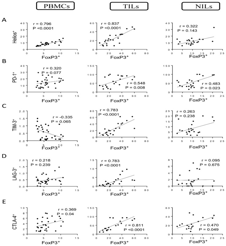
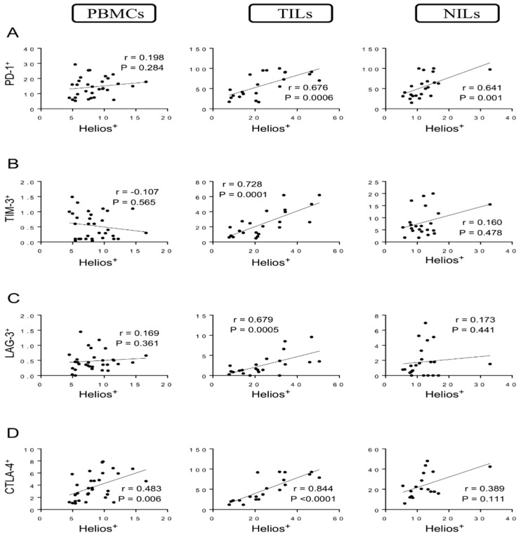
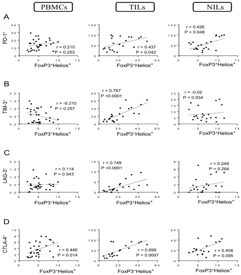
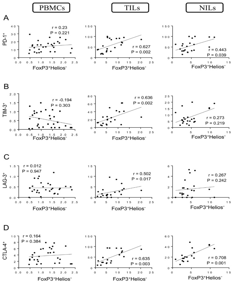
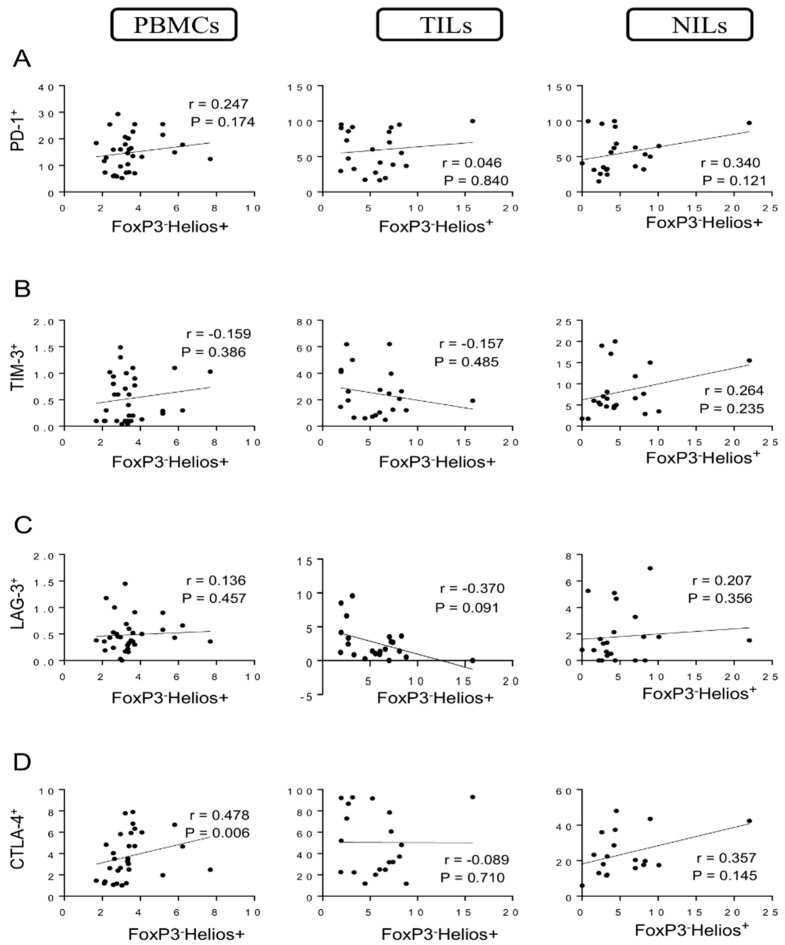
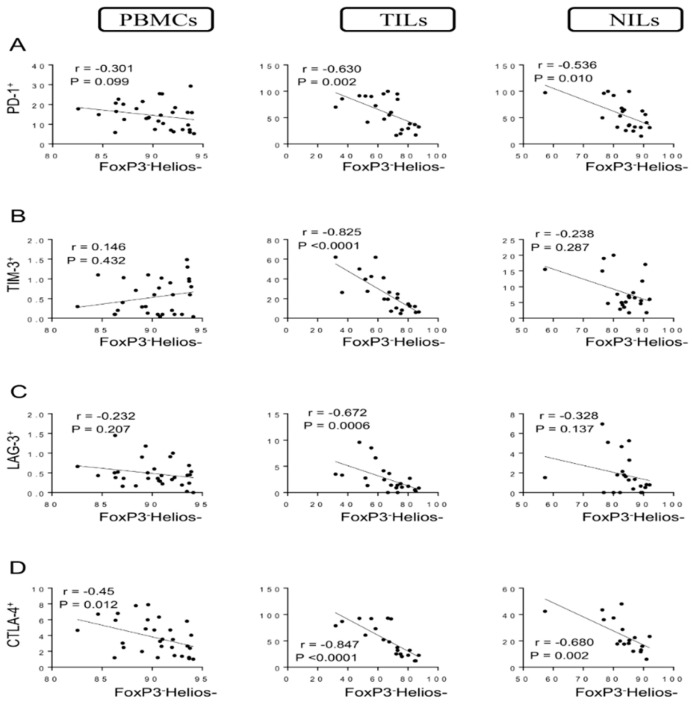
T regulatory cells (Tregs) play different roles in the regulation of anti-tumor immunity in colorectal cancer (CRC), depending on the presence of different Treg subsets. We investigated correlations between different CD4+ Treg/T cell subsets in CRC patients with immune checkpoint-expressing CD4+ T cells. Positive correlations were observed between levels of different immune checkpoint-expressing CD4+ T cells, including PD-1, TIM-3, LAG-3, and CTLA-4 with FoxP3+ Tregs, Helios+ T cells, FoxP3+Helios+ Tregs, and FoxP3+Helios- Tregs in the tumor microenvironment (TME). However, negative correlations were observed between levels of these immune checkpoint-expressing CD4+ T with FoxP3-Helios- T cells in the TME. These correlations in the TME highlight the role of cancer cells in the upregulation of IC-expressing Tregs. Additionally, positive correlations were observed between levels of FoxP3+ Tregs, Helios+ T cells, FoxP3+Helios+ Tregs, and FoxP3+Helios- Tregs and levels of CD4+CTLA-4+ T cells and CD4+PD-1+ T cells in peripheral blood mononuclear cells (PBMCs) and normal tissue-infiltrating lymphocytes (NILs). These observations suggest that CTLA-4 and PD-1 expressions on CD4+ T cell subsets are not induced only by the TME. This is the first study to investigate the correlations of different FoxP3+/-Helios+/- T cell subsets with immune checkpoint-expressing CD4+ T cells in CRC patients. Our data demonstrated strong correlations between FoxP3+/Helios+/- Tregs but not FoxP3-Helios+/- non-Tregs and multiple immune checkpoints, especially in the TME, providing a rationale for targeting these cells with highly immunosuppressive characteristics. Understanding the correlations between different immune checkpoints and Treg/T cell subsets in cancer patients could improve our knowledge of the underlying mechanisms of Treg-mediated immunosuppression in cancer.
Keywords: CRC; FoxP3; Helios; Tregs; correlation; immune checkpoints.
Conflict of interest statement
The authors declare no conflict of interest.
Figures
Similar articles
-
Correlations between Circulating and Tumor-Infiltrating CD4+ Treg Subsets with Immune Checkpoints in Colorectal Cancer Patients with Early and Advanced Stages.Vaccines (Basel). 2022 Sep 5;10(9):1471. doi: 10.3390/vaccines10091471. Vaccines (Basel). 2022. PMID: 36146549 Free PMC article.
-
Immune Checkpoints in Circulating and Tumor-Infiltrating CD4+ T Cell Subsets in Colorectal Cancer Patients.Front Immunol. 2019 Dec 17;10:2936. doi: 10.3389/fimmu.2019.02936. eCollection 2019. Front Immunol. 2019. PMID: 31921188 Free PMC article. Clinical Trial.
-
Associations of different immune checkpoints-expressing CD4+ Treg/ T cell subsets with disease-free survival in colorectal cancer patients.BMC Cancer. 2022 Jun 2;22(1):601. doi: 10.1186/s12885-022-09710-1. BMC Cancer. 2022. PMID: 35655158 Free PMC article.
-
Coexpression of Helios in Foxp3+ Regulatory T Cells and Its Role in Human Disease.Dis Markers. 2021 Jun 22;2021:5574472. doi: 10.1155/2021/5574472. eCollection 2021. Dis Markers. 2021. PMID: 34257746 Free PMC article. Review.
-
Immune checkpoint inhibitors in cancer therapy: a focus on T-regulatory cells.Immunol Cell Biol. 2018 Jan;96(1):21-33. doi: 10.1111/imcb.1003. Epub 2017 Nov 17. Immunol Cell Biol. 2018. PMID: 29359507 Review.
Cited by
-
ALDH2 as an immunological and prognostic biomarker: Insights from pan-cancer analysis.Medicine (Baltimore). 2024 Apr 19;103(16):e37820. doi: 10.1097/MD.0000000000037820. Medicine (Baltimore). 2024. PMID: 38640328 Free PMC article.
-
Correlations between Circulating and Tumor-Infiltrating CD4+ Treg Subsets with Immune Checkpoints in Colorectal Cancer Patients with Early and Advanced Stages.Vaccines (Basel). 2022 Sep 5;10(9):1471. doi: 10.3390/vaccines10091471. Vaccines (Basel). 2022. PMID: 36146549 Free PMC article.
-
Integration of peripheral blood- and tissue-based biomarkers of response to immune checkpoint blockade in urothelial carcinoma.J Pathol. 2023 Nov;261(3):349-360. doi: 10.1002/path.6197. Epub 2023 Sep 5. J Pathol. 2023. PMID: 37667855 Free PMC article.
References
LinkOut - more resources
Full Text Sources
Research Materials
