Review
. 2022 Apr 13;10(4):608.
doi: 10.3390/vaccines10040608.

COVID-19 Vaccines: Current and Future Perspectives

Affiliations
Review

COVID-19 Vaccines: Current and Future Perspectives

Luca Soraci et al. Vaccines (Basel). .

Abstract

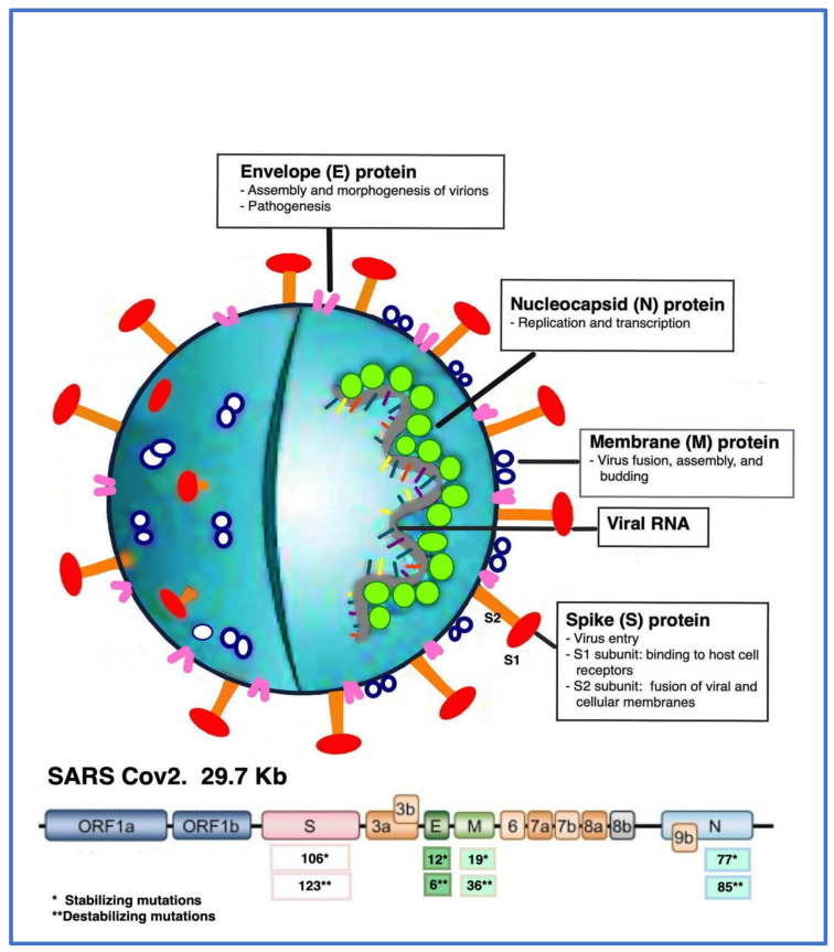
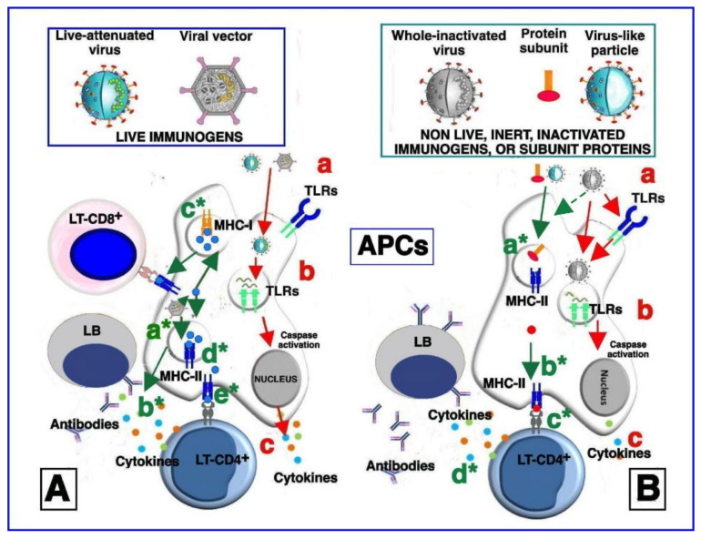
Currently available vaccines against severe acute respiratory syndrome coronavirus-2 (SARS-CoV-2) are highly effective but not able to keep the coronavirus disease 2019 (COVID-19) pandemic completely under control. Alternative R&D strategies are required to induce a long-lasting immunological response and to reduce adverse events as well as to favor rapid development and large-scale production. Several technological platforms have been used to develop COVID-19 vaccines, including inactivated viruses, recombinant proteins, DNA- and RNA-based vaccines, virus-vectored vaccines, and virus-like particles. In general, mRNA vaccines, protein-based vaccines, and vectored vaccines have shown a high level of protection against COVID-19. However, the mutation-prone nature of the spike (S) protein affects long-lasting vaccine protection and its effectiveness, and vaccinated people can become infected with new variants, also showing high virus levels. In addition, adverse effects may occur, some of them related to the interaction of the S protein with the angiotensin-converting enzyme 2 (ACE-2). Thus, there are some concerns that need to be addressed and challenges regarding logistic problems, such as strict storage at low temperatures for some vaccines. In this review, we discuss the limits of vaccines developed against COVID-19 and possible innovative approaches.

Keywords: COVID-19; S protein; innovative approaches; pandemic; recombinant vaccines.


Conflict of interest statement

The authors declare no conflict of interest.

Figures

Figure 1
Figure 1
Schematic representation of structure and genome of SARS-CoV-2. The single-stranded RNA (ssRNA) of 29.7 Kb, with nucleocapsid (N) proteins, is covered with the envelope (E) and membrane (M) proteins, whereas the spike (S) proteins are located on the outside of the viral particle. The genome is also characterized by the presence of ORFs 1a and 1b, encoding nonstructural polyproteins (NSPs), and ORFs 3a, 3b, 6, 7a, 7b, 8a, 8b, and 9b, encoding accessory proteins. The main functions of structural proteins are also indicated as follows: E protein—virion assembly, morphogenesis, and pathogenesis; N protein—genome replication and transcription; M protein—virion fusion, assembly, and budding; S protein—virus entry, with S1 subunit binding to host cell receptors (RBDs) and S2 subunit fusing viral and cellular membranes. The numbers of stabilizing and destabilizing mutations identified in the different structural proteins of SARS-CoV-2 are also indicated.
Figure 2
Figure 2
Simplified model of vaccine stimulation of the immune system. (A) Immune stimulation by live-attenuated viruses and/or vectored viral immunogens. After immunization, live-attenuated and vectored viral particles are endocytosed by antigen-presenting cells (APCs). The live-attenuated viruses can activate cell membrane pattern recognition receptors (PRRs) such as Toll-like receptors (TLRs) 2 and 6 (a). Upon entry, the live-attenuated viruses expose the nucleic acid and transcribe their genes, which, in turn, are sensed by endosomal TLRs (b). Activation of TLRs initiates signaling pathways culminating in the caspase pathway activation and production of pro-inflammatory and antiviral cytokines and chemokines (c). Immune stimulation by vectored viral immunogens may induce, via NOD-like receptor family pyrin domain-containing (NLRP) 3 pathway, inflammasome activation (a*) and cytokine production (b*). The transcribed vector-encoded transgene generates the immunogenic proteins (blue circles), which can then be proteosome-processed and associated with class I major histocompatibility complex (MHC-I) (c*) or with class II major histocompatibility complex (MHC-II) in endocytic vesicles (d*). MHC-I molecules loaded with transgenic epitopes translocate to the cell membrane, where they are recognized by antigen-specific CD8+ T cells (e*). Consequently, the infected cell is killed, and releases antigens in the extracellular space. In the same way, MHC-II molecules loaded with transgenic epitopes and translocated to the cell membrane are recognized by CD4+ helper T cells, which secrete cytokines and chemokines and further activate antigen-specific CD8+ T cells and B cells. Finally, stimulated B cells maturate into antibody-secreting plasma cells and/or memory B cells, as well as a portion of the stimulated T cells, which become memory cells (not shown). Overall, live immunogens are able to equally stimulate both humoral and cell-associated immune responses). (B) Immune stimulation by non-live (inert) inactivated vaccines, protein subunits, and virus-like particles. Immunization with antigens, inoculated together with the adjuvants that are added to vaccine formulations induces cytokine production from local cells. Cytokines in turn activate and/or attract APCs to the immunization site. In addition, the antigens may directly activate APCs through binding to cell membrane TLRs (a). The inactivated viruses are phagocytized by APCs and nucleic-acid traces inside the phagosomes and may activate endosomal TLRs (b), leading to the production of cytokines and chemokines (in a smaller amount, compared to (A) (c). The antigens contained in the inert vaccines in protein subunit formulations and in virus-like particles, after the entry in the call, are also degraded inside the endocytic vesicles, loaded onto MHC-II molecules (a*), and presented to CD4+ T cells (b*). Activation of CD4+ T lymphocytes leads to production of cytokines and chemokines, which induce the activation of antigen-specific B cells (c*), which maturate into antibody-secreting plasma cells (d*) and/or memory B cells. In general, inert antigens, such as nonlive inactivated vaccines, protein subunits, and virus-like particles, induce potent humoral responses and low-to-moderate T-cell responses. Activation of CD8+ T cells by inert antigens occurs through alternative pathways that are not depicted in this figure. The stimulation processes described and depicted in steps (a), (b), and (c) is reported less frequently and is less potent than stimulation by live immunogens (A), as depicted in (B) panel).
Figure 3
Figure 3
Comparative analysis of adverse reactions related to ACE2 following SARS-CoV-2 infection (A) and SARS-CoV-2 mRNA vaccination (B). (A) SARS-CoV-2 stimulates innate immune cells through both exogenous and endogenous TLR ligands (1a), inducing a strong TLR signaling that leads to cytokine storm (2a) which then induces a cascade of adverse events, including arterial thrombosis, venous thromboembolism, pulmonary edema, and interstitial inflammation * (3a). In addition, the S protein binds both to the ACE2 receptor and to soluble ACE2 (1b). S protein also induces Ab1 production (3b). Ab1, in turn, induces anti-idiotypic Ab2 production, which is able to bind to RBD of the anti-S protein antibodies as well as to both soluble ACE2 and ACE2 receptors, causing specific inhibition (3b). (B) Liposomal nanoparticles containing mRNA encoding viral S protein are phagocytized by innate immune cells (1a) and mRNA binds to specific endogenous TLRs. At the same time, mRNA molecules are translated into S proteins inside the immune cells that can then present them in association with HLA class II antigens, constitutively expressed on professional antigen-presenting cells, such as dendritic cells and monocytes/macrophages (1b). The binding of mRNA to endogenous TLRs induces more attenuated cytokine production (2a), which could lead to adverse events similar to those induced by SARS-CoV-2 infection, despite, in this case, being more attenuated and much less common (see the colored text boxes in lighter red, compared with red boxes in (A). The S protein presentation induces both humoral (2b) and cellular immune responses. Anti-idiotypic Ab2 production, with consequent inhibition of ACE2 activity, is also more attenuated compared with SARS-CoV2 infection).

References

    1. Frutos R., Serra-Cobo J., Pinault L., Lopez Roig M., Devaux C.A. Emergence of Bat-Related Betacoronaviruses: Hazard and Risks. Front. Microbiol. 2021;12:591535. doi: 10.3389/fmicb.2021.591535. - DOI - PMC - PubMed
    1. Johnson N.P., Mueller J. Updating the accounts: Global mortality of the 1918–1920 “Spanish” influenza pandemic. Bull. Hist. Med. 2002;76:105–115. doi: 10.1353/bhm.2002.0022. - DOI - PubMed
    1. Khubchandani J., Jordan T.R., Yang Y.T. Ebola, Zika, Corona… What Is Next for Our World? Int. J. Environ. Res. Public Health. 2020;17:3171. doi: 10.3390/ijerph17093171. - DOI - PMC - PubMed
    1. Giannotta G., Giannotta N. mRNA COVID-19 Vaccines and Long-Lived Plasma Cells: A Complicated Relationship. Vaccines. 2021;9:1503. doi: 10.3390/vaccines9121503. - DOI - PMC - PubMed
    1. Mistry P., Barmania F., Mellet J., Peta K., Strydom A., Viljoen I.M., James W., Gordon S., Pepper M.S. SARS-CoV-2 Variants, Vaccines, and Host Immunity. Front. Immunol. 2021;12:809244. doi: 10.3389/fimmu.2021.809244. - DOI - PMC - PubMed

LinkOut - more resources