. 2022 Apr 14;10(4):615.
doi: 10.3390/vaccines10040615.

Mycobacterium bovis Wild-Type BCG or Recombinant BCG Secreting Murine IL-18 (rBCG/IL-18) Strains in Driving Immune Responses in Immunocompetent or Immunosuppressed Mice

Affiliations

Mycobacterium bovis Wild-Type BCG or Recombinant BCG Secreting Murine IL-18 (rBCG/IL-18) Strains in Driving Immune Responses in Immunocompetent or Immunosuppressed Mice

Marek Fol et al. Vaccines (Basel). .

Abstract

Mycobacterium tuberculosis infections remain a global health problem in immunosuppressed patients. The effectiveness of BCG (Bacillus Calmette−Guérin), an anti-tuberculosis vaccine, is unsatisfactory. Finding a new vaccine candidate is a priority. We compared numerous immune markers in BCG-susceptible C57BL/6 and BCG-resistant C3H mice who had been injected with 0.9% NaCl (control) or with wild-type BCG or recombinant BCG secreting interleukin (IL)-18 (rBCG/IL-18) and in immunized mice who were immunocompromised with cyclophosphamide (CTX). The inoculation of rBCG/IL-18 in immunocompetent mice increased the percentage of bone marrow myeloblasts and promyelocytes, which were further elevated in the rBCG/IL-18/CTX-treated mice: C57BL/6 mice—3.0% and 11.4% (control) vs. 18.6% and 42.4%, respectively; C3H mice—1.1% and 7.7% (control) vs. 18.4% and 44.9%, respectively, p < 0.05. The bone marrow cells showed an increased mean fluorescence index (MFI) in the CD34 adhesion molecules: C57BL/6 mice—4.0 × 10³ (control) vs. 6.2 × 10³; C3H mice—4.0 × 10³ (control) vs. 8.0 × 10³, p < 0.05. Even in the CTX-treated mice, the rBCG/IL-18 mobilized macrophages for phagocytosis, C57BL/6 mice—4% (control) vs. 8%; C3H mice—2% (control) vs. 6%, and in immunocompetent mice, C57BL/6 induced the spleen homing of effector memory CD4+ and CD8+ T cells (TEM), 15% (control) vs. 28% and 8% (control) vs. 22%, respectively, p < 0.05. In conclusion, rBCG/IL-18 effectively induced selected immune determinants that were maintained even in immunocompromised mice.

Keywords: BCG; C3H; C57BL/6; IL-18; cyclophosphamide; immunity; immunosuppression.


Conflict of interest statement

The authors declare no conflict of interest. The funders had no role in the design of the study; in the collection, analyses, or interpretation of data; in the writing of the manuscript; or in the decision to publish the results.

Figures
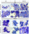
Figure 1
Figure 1
Morphology of bone marrow cells isolated from C3H and C57BL/6 mice who had been immunocompromised with cyclophosphamide or immunocompetent animals who had been injected with physiological saline (NaCl) and immunized with Mycobacterium bovis (wild-type BCG) or the recombinant BCG (rBCG) strain secreting interleukin (IL)-18. Representative pictures are shown for each group. Cytospins were prepared from freshly isolated bone marrow and stained with the May–Grünwald for 6 min and Giemsa solution for another 15 min. Finally, the slides were rinsed with water and placed to dry in air. The cells were observed using a light microscope at 100× magnification (immersion). The experiment was triplicated, and each time, at least 12 randomly selected view fields per slide were captured. The cells were identified on the basis of histological/morphological markers by two independent histopathologists from Institut für Tierpathologie, Berlin, Germany, and are indicated by arrows and numbers: (1) myelocyte, (2) promyelocyte, (3) metamyelocyte, (4) myeloblast, (5) monocyte, (6) lymphocyte, (e1) eosinophilic myelocyte, (e2) eosinophilic promyelocyte, (e3) eosinophilic metamyelocyte, (MIT) mitosis, (pE) proerythroblast, (pcE) polychromatic erythroblast, and (ocE) orthochromatic erythroblast.
Figure 2
Figure 2
The expression of CD34 and CD117 on bone marrow cells in C57BL6 and C3H mice, in immunocompetent mice and in mice who were immunocompromised with cyclophosphamide (CTX), and in mice who were non-immunized or immunized with Mycobacterium bovis wild-type BCG or by the recombinant BCG (rBCG) strain secreting IL-18. (A) Representative confocal laser scanning microscopy images (NikonD-Eclipse C1 microscope equipped with an inverted 60× objective). Bone marrow cells were stained with DyLight 488-conjugated rat monoclonal anti-mouse CD34 (B) and with allophycocyanin (APC)-conjugated rat monoclonal anti-mouse CD117 (C). Data are shown as mean ± SEM (standard error of the mean). Asterisk (*) indicates p value ≤ 0.05; NaCl—physiological saline solution NaCl 0.9%; CD—cluster of differentiation; MFI—mean fluorescence intensity.
Figure 3
Figure 3
Phagocytic activity of alveolar macrophages in C57BL6 or C3H mice who were immunocompetent or immunocompromised with cyclophosphamide (CTX) and who were non-immunized or immunized with Mycobacterium bovis wild-type BCG or the recombinant BCG strain (rBCG) secreting IL-18. The percentage of macrophages with at least one engulfed bacterium (A). The mean number of bacteria that were phagocytized by one macrophage (B). At least three hundred of alveolar macrophages were counted in each slide. The number of ingested bacteria and the number of phagocytes containing at least one bacterium were determined. The percentage of macrophages engaged in phagocytosis (with at least 1 bacterium) was calculated as follows: (number of phagocytes containing bacteria/total number of phagocytes counted in slide) × 100%. The mean number of bacteria phagocytized by one macrophage was calculated as follows: total number of ingested bacteria/number of phagocytes containing at least one bacterium. Data are shown as mean ± SEM (standard error of the mean). Differences were calculated using a Kruskal–Wallis test, and statistical significances are indicated vs. control mice. Asterisk (*) indicates p value < 5; NaCl—physiological saline solution NaCl 0.9%.
Figure 4
Figure 4
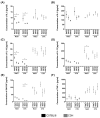
Serum concentrations of the studied cytokines in the sera from C57BL/6 and C3H mice who had been immunized with wild-type BCG or the recombinant BCG strain (rBCG) secreting IL-18 and who were immunocompetent or cyclophosphamide (CTX)-compromised. Data are shown as mean ± SEM (standard error of the mean). For each condition, samples were tested. p values were calculated using a one-way analysis of variance (ANOVA). IL-2 (A), IL-5 (B), IL-10 (C), IL-12 (D), granulocyte macrophage colony stimulating factor (GM-CSF) (E), tumor necrosis factor alpha (TNF)-α (F). Black dots—C57BL/6 mice; grey dots—C3H mice. Asterisk (*) indicates p value ≤ 0.05.
Figure 5
Figure 5
Comparison of central (TCM) and effector memory T cells (TEM) in C57BL6 and C3H mice who were non-immunized or immunized with the wild-type BCG or recombinant BCG (rBCG) secreting IL-18 and in mice who were immunocompromised with cyclophosphamide (CTX) or who were immunocompetent (NaCl, Control). Graphs show cumulative data for n = 7 mice/group. Data are expressed as mean ± SD (standard deviation) TCM or TEM percentages; * p value < 0.05, ** p value < 0.01. (A) total number of CD4+ T cells, (B) total number of CD8+ T cells, (C) number of CD4+ TCM cells, (D) number of CD8+ TCM cells, (E) number of CD4+ TEM cells, (F) number of CD8+ TEM cells.

Similar articles

References

    1. Global Tuberculosis Report WHO 2021. [(accessed on 17 February 2022)]. Available online: https://www.who.int/teams/global-tuberculosis-programme/tb-reports.
    1. Houben R.M., Dodd P.J. The global burden of latent tuberculosis infection: A re-estimation using mathematical modelling. PLoS Med. 2016;13:e1002152. doi: 10.1371/journal.pmed.1002152. - DOI - PMC - PubMed
    1. Kiazyk S., Ball T.B. Latent tuberculosis infection: An overview. Can. Commun. Dis. Rep. 2017;43:62–66. doi: 10.14745/ccdr.v43i34a01. - DOI - PMC - PubMed
    1. Ai J.W., Ruan Q.L., Liu Q.H., Zhang W.H. Updates on the risk factors for latent tuberculosis reactivation and their managements. Emerg. Microbes Infect. 2016;5:e10. doi: 10.1038/emi.2016.10. - DOI - PMC - PubMed
    1. Muñoz L., Stagg H.R., Abubakar I. Diagnosis and management of latent tuberculosis infection. Cold Spring Harb. Perspect. Med. 2015;5:a017830. doi: 10.1101/cshperspect.a017830. - DOI - PMC - PubMed