. 2022 Apr 15;10(4):620.
doi: 10.3390/vaccines10040620.

Adjuvant Curdlan Contributes to Immunization against Cryptococcus gattii Infection in a Mouse Strain-Specific Manner

Affiliations

Adjuvant Curdlan Contributes to Immunization against Cryptococcus gattii Infection in a Mouse Strain-Specific Manner

Patrícia Kellen Martins Oliveira-Brito et al. Vaccines (Basel). .

Abstract

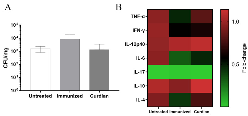
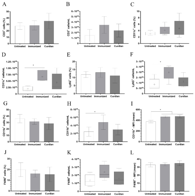
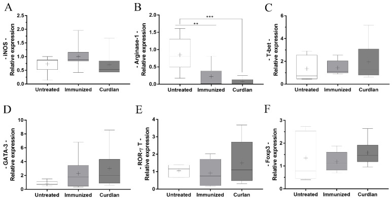
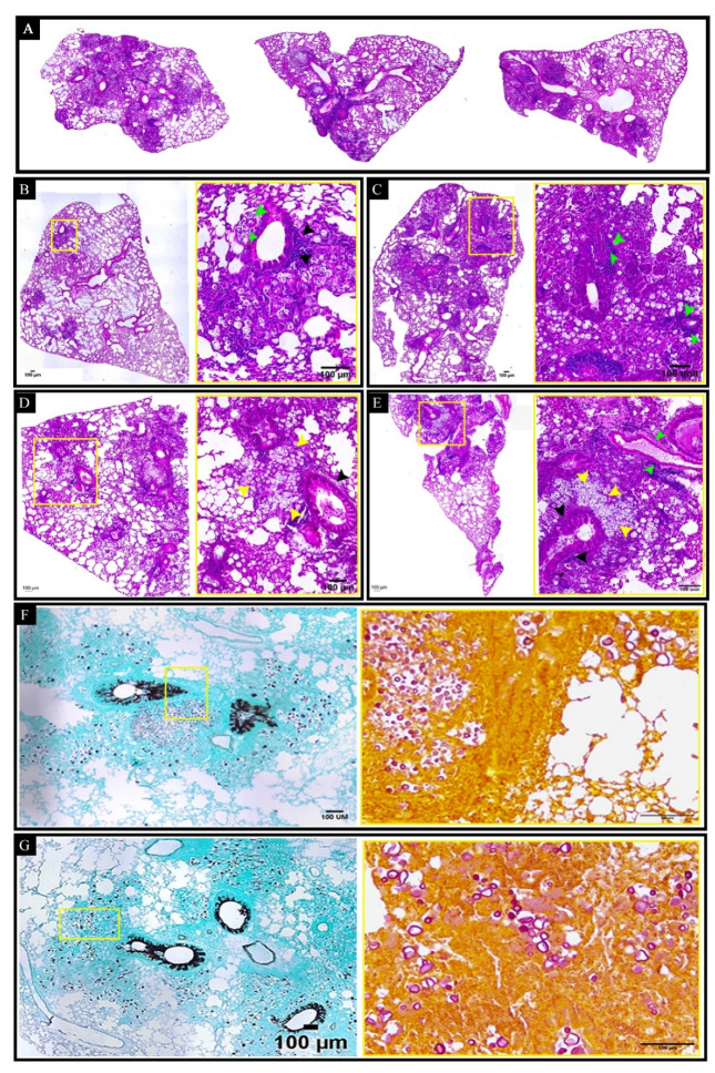
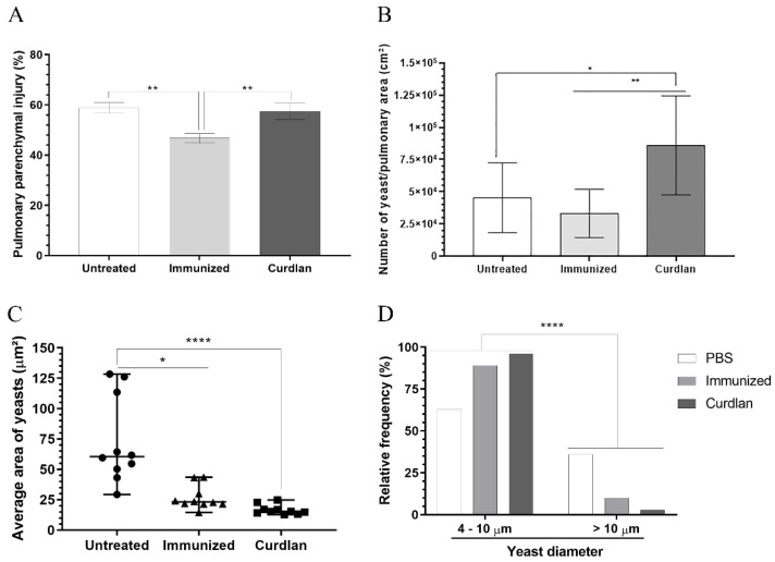
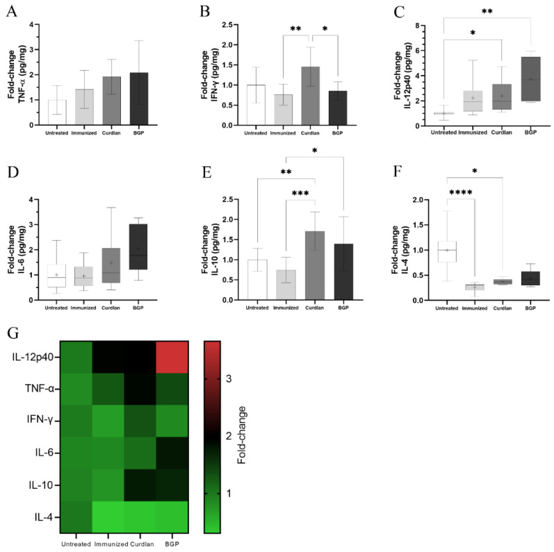
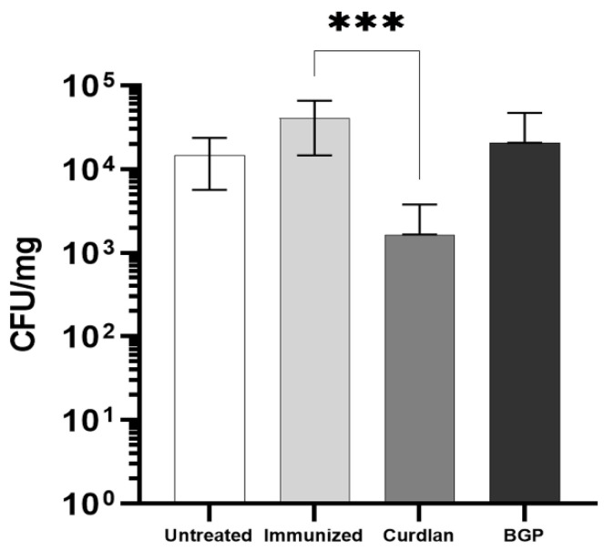
The low efficacy and side effects associated with antifungal agents have highlighted the importance of developing immunotherapeutic approaches to treat Cryptococcus gattii infection. We developed an immunization strategy that uses selective Dectin-1 agonist as an adjuvant. BALB/c or C57BL/6 mice received curdlan or β-glucan peptide (BGP) before immunization with heat-killed C. gattii, and the mice were infected with viable C. gattii on day 14 post immunization and euthanized 14 days after infection. Adjuvant curdlan restored pulmonary tumor necrosis factor- α (TNF-α) levels, as induced by immunization with heat-killed C. gattii. The average area and relative frequency of C. gattii titan cells in the lungs of curdlan-treated BALB/c mice were reduced. However, this did not reduce the pulmonary fungal burden or decrease the i0,nflammatory infiltrate in the pulmonary parenchyma of BALB/c mice. Conversely, adjuvant curdlan induced high levels of interferon-γ (IFN-γ) and interleukin (IL)-10 and decreased the C. gattii burden in the lungs of C57BL/6 mice, which was not replicated in β-glucan peptide-treated mice. The adjuvant curdlan favors the control of C. gattii infection depending on the immune response profile of the mouse strain. This study will have implications for developing new immunotherapeutic approaches to treat C. gattii infection.

Keywords: Cryptococcus gattii; Dectin-1; curdlan; immunotherapy; β-glucan peptide.


Conflict of interest statement

The authors declare no conflict of interest.

Figures

Figure 1
Figure 1
Evaluation of the immunotherapeutic strategy combined with the adjuvant curdlan against C. gattii challenge. (A) BALB/c mice were intraperitoneally (i.p.) administered curdlan (200 μg/mouse) or phosphate-buffered saline (PBS) on day 0. On day 3, they were immunized with 2 × 10⁷ yeast cells of heat-inactivated Cryptococcus gattii (HK-C. gattii-thin) or administered PBS intranasally (i.n.), and booster doses were administered after 14 days. (B,C) On days 38 and 45 (7 and 14 days post immunization (d.p.i.), respectively), the levels of serum immunoglobulin M (IgM) and IgG anti-glucuronoxylomannan (GXM) antibodies were measured (dilutions of 1:200). On day 45, the mice were i.n.-infected with 1 × 10⁵ C. gattii yeast cells. (D) On day 59 (14 days post infection), the mice were euthanized and their serum titrated to measure the IgM and IgG anti-GXM levels (IgM dilutions of 1:10, 1:20, 1:40, 1:80, and 1:160; and IgG anti-GXM dilutions of 1:50, 1:100, 1:200, 1:400, and 1:800). The results are expressed as mean ± SD. * p < 0.05.
Figure 2
Figure 2
Isotyping of serum immunoglobulins in BALB/c mice and the effect of serum on the growth curve of C. gattii cultivated in titan cell medium (TCM). BALB/c mice immunostimulated with curdlan and/or immunized with HK-C. gattii-thin were euthanized 14 days post infection with C. gattii. The serum was collected and used for both isotyping the immunoglobulins and incubating with C. gattii to determine its growth curve. The (A) IgG1, (B) IgG2a, (C) IgG2b, (D) IgG3, (E) IgM, (F) IgA isotypes, and the (G) kappa and (H) lambda light chains were evaluated. (I) C. gattii R265 was cultivated at 37 °C for 24 h in TCM, and the growth curves were determined in the presence of serum from mice belonging to the untreated (black circle), immunized (red square), and curdlan groups (blue triangle). The results are expressed as mean ± SD. * p < 0.05, ** p < 0.01, and *** p < 0.001.
Figure 3
Figure 3
Evaluation of the size and concentration of C. gattii cultured in TCM in the presence of serum from BALB/c mice treated with the adjuvant curdlan. The BALB/c mice received curdlan or PBS and were immunized with HK-C. gattii-thin, before being challenged with C. gattii. After 14 days of infection, the serum was collected and incubated with C. gattii cultured in TCM. After 24 h of incubation, the C. gattii cells were analyzed by flow cytometry. The size and concentration of the three populations of C. gattii cells, as delimited by gates 1 (red; (A,B)), 2 (blue; (A,C)), and 3 (pink; (A,D)), were measured. The FSC-HLin was the parameter used to normalize the size among the groups to express the cell concentration (cell/mL). Results are expressed as medians with interquartile ranges.
Figure 4
Figure 4
Measurement of the pulmonary fungal burden and cytokine levels in the lungs of immunized BALB/c mice after C. gattii challenge. After administering curdlan or PBS, the mice were immunized with HK-C. gattii-thin. On day 45 (14 d.p.i.), the mice were challenged with C. gattii. On day 59 (14 d.p.i.), the lungs of the mice were harvested and homogenized. The supernatants were used to (A) determine the colony-forming units (CFU), normalized to the lung mass (CFU/mg), and (B) measure the levels of cytokines tumor necrosis factor-α (TNF-α), interferon-γ (IFN-γ), interleukin (IL)-12p40, IL-6, IL-17, IL-10, and IL-4 using enzyme-linked immunosorbent assay (ELISA). Values are expressed as mean ± SD or as medians with maximum and minimum values.
Figure 5
Figure 5
Phenotyping of the leukocytes in the lungs of mice treated with curdlan and immunized with HK-C. gattii. After administration of curdlan or PBS, the mice were immunized with HK-C. gattii-thin and infected with viable C. gattii. The lungs of the infected mice were harvested at 14 days post infection. Pulmonary leukocytes were obtained by dissociating the tissue with collagenase. The cell suspensions were used to determine the frequency (%) and concentration (cell/mL) of T lymphocytes (A,B), CD11c+ (C,D), Ly6G+ (E,F), CD11b+ (G,H), and F4/80+ (J,K). The mean fluorescence intensity (MFI) of (I) CD11b+ and (L) F4/80+ cells was recorded using flow cytometry. The relative and absolute frequencies of these cells were determined in the lung samples. The results are expressed as both, in percentage and cells/mL, and are represented as mean ± SD or as median with maximum and minimum values. Comparisons were made between groups for the same period of infection. * p < 0.05.
Figure 6
Figure 6
Relative expression of transcription factors related to macrophage polarization and T-helper cell differentiation in the lungs of BALB/c mice treated with curdlan and immunized with HK-C. gattii. Lungs were harvested from the mice 14 d.p.i., and total RNA was extracted from the lungs. The total RNA was reverse-transcribed into complementary DNA (cDNA), and the relative expression levels of (A) iNOS, (B) Arginase-1, (C) T-bet, (D) GATA-3, (E) ROR-γt, and (F) FOXP3 were determined by real-time quantitative polymerase chain reaction (qRT-PCR). The values were normalized to β-actin expression. The results are expressed as median, maximum, and minimum values. Comparisons were made between groups for the same period of infection. ** p < 0.01, and *** p < 0.001.
Figure 7
Figure 7
Histopathological analysis of the lungs of BALB/c mice treated with curdlan and immunized with HK-C. gattii. (A) The panels show representative lung sections from untreated mice (left side) or those that were immunized with HK-C. gattii in the presence (right size) or absence (middle size) of curdlan. (BE) The total lung (10× magnification) and lung sections (40× magnification) have been shown for each panel. The sections were stained with hematoxylin and eosin (H&E). Perivasculitis (green arrows) and peribrochiolitis (black arrows) were observed, with exudates primarily composed of mononuclear leukocytes. Inflammatory infiltrate composed of mononuclear leukocytes and neutrophils filling the alveolar space contained oval to rounded (yellow arrows) yeast cells diffusely distributed or restricted to the vacuolated cytoplasm of macrophages. The nodules consisted of high concentrations of aggregated C. gattii yeasts (yellow arrows). (F,G) The panels show representative lung sections for the different groups. The sections were stained with Grocott (left side, 10× magnification) and Mucicarmin (right side; 20× magnification) stains to visualize the distribution of fungi. The images presented are representative fields of the different groups. Five mice were analyzed per group. Magnification bar: 100 µm.
Figure 8
Figure 8
Morphometric analysis of the lung tissue in BALB/c mice treated with curdlan, immunized with HK-C. gattii, and infected with viable C. gattii. Lungs were perfused with PBS and harvested from the BALB/c mice 14 days post infection. Lung fragments were fixed with methacarn, dehydrated in an alcohol battery, diaphanized with xylol, and embedded in paraffin. Serial 5 µm cuts were made and stained with Grocott-Gomori methenamine silver (GMS). (A) Pulmonary parenchyma injury (%), (B) quantification of the number of yeast cells/pulmonary area (cm2), (C) average yeast cell area (µm2), and (D) yeast relative frequency were evaluated in a semi-automated quantitative way using ImageJ Fiji. The data were analyzed using the X2 test (Goodness-of-fit test). Values were expressed as mean ± SD. * p < 0.05, ** p < 0.01, and **** p < 0.0001.
Figure 9
Figure 9
The levels of IgM and IgG anti-GXM antibodies in the serum and growth curve for C. gattii after incubation with serum from immunized C57BL/6 mice. After administering Dectin-1 ligands or PBS, C57BL/6 mice were immunized with HK-C. gattii-thin. On day 45 (14 d.p.i.), the mice were challenged with C. gattii. On day 59 (14 d.p.i.), mice serum and lungs were collected. Several dilutions of the serum were used to measure the levels of IgM and IgG anti-GXM antibodies (IgM = 1:10, 1:20, 1:40, 1:80, 1:160, 1:320, and 1:640; IgG = 1:50, 1:100, 1:200, 1:400, 1:800, 1:1600, and 1:3200).
Figure 10
Figure 10
Measurement of cytokine levels in the lungs of immunized C57BL/6 mice challenged with C. gattii. After the administration of curdlan, β-glucan peptide (BGP), or PBS, C57BL/6 mice were immunized with HK-C. gattii-thin. On day 45 (14 d.p.i.), the mice were challenged with C. gattii. On day 59 (14 days post infection), mice lungs were harvested and homogenized, and the supernatants were used for measuring the levels of (A) TNF-α, (B) IFN-γ, (C) IL-12p40, (D) IL-6, (E) IL-10, and (F) IL-4 cytokines via ELISA. (G) Fold change (1–3 times) in the cytokine levels in all groups (in comparison to the untreated group) are represented in the heat map. Results are expressed as mean ± SD or as medians with interquartile ranges. * p < 0.05, ** p < 0.01, *** p < 0.001, and **** p < 0.0001.
Figure 11
Figure 11
Quantification of the pulmonary fungal burden in C57BL/6 mice subjected to the HK-C. gattii immunization protocol, with or without the administration of curdlan. C57BL/6 mice received curdlan (200 μg/mouse), BGP (200 μg/mouse), or PBS i.p. on day 0. On days 3, 17, and 31, they were immunized with 2 × 10⁷ HK-C. gattii-thin yeast cells or administered PBS i.n., with an interval of 14 days between immunizations. On day 45 (14 d.p.i.), the mice were infected with C. gattii. On day 59 (14 days post infection), the mice were euthanized, and the pulmonary fungal burden was quantified using the CFU (colony-forming units) assay and normalized to the lung mass (CFU/mg). The results are expressed as mean ± SD. *** p < 0.001.
Figure 12
Figure 12
Evaluation of the growth and concentration of C. gattii cells cultured in TCM and incubated with serum from C57BL/6 mice treated with the Dectin-1 ligand. C57BL/6 mice that had received curdlan, BGP, or PBS were immunized with HK-C. gattii-thin before being challenged with C. gattii. Serum was collected after 14 days of infection and incubated with C. gattii in TCM. (A) C. gattii R265 was cultured at 37 °C for 18, 21, and 24 h in TCM, and the growth curves were determined in the presence of serum from untreated (black circle), immunized (red square), curdlan (blue triangle), and BGP groups (green triangle). (BE) After 24 h of incubation, C. gattii was analyzed by flow cytometry. The cell size and concentration of the three populations as defined by gates 1 (red; (B,C)), 2 (blue; (B,D)), and 3 (pink; (B,E)) were measured. Cell concentration is expressed in cell/mL. Results are expressed as mean ± SD, or as medians, and maximum and minimum values. * p < 0.05.

Similar articles

Cited by

References

    1. Jermy A. Stop neglecting fungi. Nat. Microbiol. 2017;2:17120. doi: 10.1038/nmicrobiol.2017.120. - DOI - PubMed
    1. Hadrich I., Ayadi A. Epidemiology of antifungal susceptibility: Review of literature. J. Mycol. Med. 2018;28:574–584. doi: 10.1016/j.mycmed.2018.04.011. - DOI - PubMed
    1. Denning D.W. Calling upon all public health mycologists. Eur. J. Clin. Microbiol. Infect. Dis. 2017;36:923–924. doi: 10.1007/s10096-017-2909-8. - DOI - PubMed
    1. Bongomin F., Gago S., Oladele R., Denning D. Global and multi-national prevalence of fungal diseases—estimate precision. J. Fungi. 2017;3:57. doi: 10.3390/jof3040057. - DOI - PMC - PubMed
    1. Dixit A., Carroll S.F., Qureshi S.T. Cryptococcus gattii: An emerging cause of fungal disease in North America. Interdiscip. Perspect. Infect. Dis. 2009;2009:840452. doi: 10.1155/2009/840452. - DOI - PMC - PubMed

LinkOut - more resources