Review
. 2022 Mar 25;15(4):401.
doi: 10.3390/ph15040401.

Regulation of the Homeostatic Unfolded Protein Response in Diabetic Nephropathy

Affiliations
Review

Regulation of the Homeostatic Unfolded Protein Response in Diabetic Nephropathy

Hongjie Wang et al. Pharmaceuticals (Basel). .

Abstract

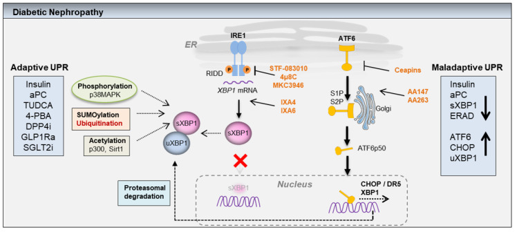
A growing body of scientific evidence indicates that protein homeostasis, also designated as proteostasis, is causatively linked to chronic diabetic nephropathy (DN). Experimental studies have demonstrated that the insulin signaling in podocytes maintain the homeostatic unfolded protein response (UPR). Insulin signaling via the insulin receptor non-canonically activates the spliced X-box binding protein-1 (sXBP1), a highly conserved endoplasmic reticulum (ER) transcription factor, which regulates the expression of genes that control proteostasis. Defective insulin signaling in mouse models of diabetes or the genetic disruption of the insulin signaling pathway in podocytes propagates hyperglycemia induced maladaptive UPR and DN. Insulin resistance in podocytes specifically promotes activating transcription factor 6 (ATF6) dependent pathogenic UPR. Akin to insulin, recent studies have identified that the cytoprotective effect of anticoagulant serine protease-activated protein C (aPC) in DN is mediated by sXBP1. In mouse models of DN, treatment with chemical chaperones that improve protein folding provides an additional benefit on top of currently used ACE inhibitors. Understanding the molecular mechanisms that transmute renal cell specific adaptive responses and that deteriorate renal function in diabetes will enable researchers to develop new therapeutic regimens for DN. Within this review, we focus on the current understanding of homeostatic mechanisms by which UPR is regulated in DN.

Keywords: ATF6; ER stress; XBP1; aPC; diabetic nephropathy; insulin signaling; podocytes; unfolded protein response.


Conflict of interest statement

The authors declare no conflict of interest.

Figures

Figure 1
Figure 1
(a,b): Scheme showing adaptive (homeostatic) and maladaptive unfolded protein response (UPR) in diabetic nephropathy (DN). Adaptive UPR: Accumulation of misfolded proteins within the ER causes ER stress, which is sensed by the three major ER-transmembrane proteins IRE1, PERK, and ATF6. IRE1 pathway: Upon activation, IRE1 through its endoribonuclease activity induces the splicing of ER-transcription factor XBP1. The spliced XBP1 (sXBP1) translocates to the nucleus and induces the expression of genes that encode proteins involved in mitigation of ER stress. Independent of IRE1 activation, the nuclear translocation of sXBP1 is mediated by insulin and coagulation protease activated protein C (aPC). Within the insulin sensitive renal podocytes, insulin signaling via insulin receptor and the downstream regulatory subunits of PI3Kinase, p85α, and p85β promotes nuclear translocation of sXBP1. Akin to insulin, aPC signaling via protease activated receptors promotes nuclear translocation of sXBP1 in a p85α and a p85β dependent fashion. Both insulin and aPC signaling in glomerular cell types (podocytes and endothelial cells) specifically activates sXBP1 dependent proteostasis pathway, without inducing PERK or ATF6 pathways. During severe or unmitigated ER stress, IRE1α-RNase can also cleave ER-localized mRNAs or non-coding functional RNAs, leading to their degradation through regulated IRE1-dependent decay (RIDD). RIDD not only reduces the ER protein folding load, but also modulates inflammation, metabolism, and inflammasome signaling pathway. Additionally, the cytoplasmic domain of IRE1α serves as a scaffold, which recruits adaptor proteins (e.g., tumor necrosis factor receptor-associated factor (TRAF). The IRE1/TRAF complexes activate inflammatory responses under non-canonical ER stress conditions. PERK pathway: Upon activation, PERK induces the phosphorylation of eIF2α which transiently attenuates the translation in order to reduce protein synthesis. Concomitant activation of stress inducible transcription factor ATF4 induces the expression of genes that are involved in redox homeostasis, amino acid metabolism, autophagy, ER-protein folding, and apoptosis. Additionally, ATF4 participates in a feedback loop mediated by PPI and GADD34 to dephosphorylate eIF2α and to restore protein synthesis. However, severe ER stress may trigger ATF4-dependent cell death program by activation of CHOP. Thus, the magnitude of ER stress and the disease-specific stimuli may disparately modulate PERK-ATF4 dependent adaptive and maladaptive cellular responses. ATF6 pathway: Upon activation ATF6 relocates to the Golgi-complex. Within the Golgi-complex the full length ATF6α-p90 undergoes proteolytic cleavage mediated by site1 and site2 proteases (S1P and S2P), resulting in a highly active transcription factor ATF6α-p50, which translocates to the nucleus to induce gene expression. ATF6 induces the expression of genes that are involved in ER-protein folding, protein secretion, ERAD, autophagy, and apoptosis. Figure 1b: Maladaptive UPR in diabetic nephropathy (DN). The disease-specific stimuli (for e.g., hyperglycemia) in chronic DN coupled with loss of insulin or insulin resistance diminishes the nuclear translocation of sXBP1. Subsequently, chronic hyperglycemia specifically activates ATF6 pathway, which promotes the expression of proapoptotic CHOP and DN. Of note, activation of the PERK-ATF4 pathway was not observed in DN, suggesting cell- and disease-specific regulation of the UPR. The loss of the sXBP1 function often results in hyperactivation of IRE1α. The IRE1α-TRAF2/JNK and IRE1α-RIDD axis activates BAX/BAK mediated mitochondrial dysfunction and apoptosis by activating caspase-2 or caspase-8 dependent activation of BH3-interacting domain death agonist (BID). Additionally, the IRE1α-RIDD axis promotes degradation of microRNA-17(miR-17), which stabilizes the thioredoxin-interacting protein (TXNIP) and promotes inflammasome activation. Inflammasome activation mediated by TXNIP or mitochondrial reactive oxygen species (ROS) promotes caspase-1/IL-1β/IL-18 dependent sterile inflammation. The IRE1α-TRAF2/JNK signaling also modulates autophagy. Additionally, persistent activation of ATF6α in DN may promote a CHOP-dependent apoptosis program. CHOP promotes ER stress induced apoptosis by activating death receptor 5 (DR5) dependent caspase-8 activation or by activating the Bcl-2 and BH3-only family members NOXA, PUMA, BID, BAX, and BAK.
Figure 2
Figure 2
UPR regulating genes: Glomerular-specific gene expression in the Nephromine database (within “Ju podocyte” dataset). A large number of unfolded protein response genes are induced in human patients with well-established DN when compared to healthy controls (1: Healthy living donors (n = 41); 2: DN: diabetic nephropathy (n = 12); data queried for overexpression).
Figure 3
Figure 3
Potential mechanisms that modulate UPR in diabetic nephropathy (DN): post-translational modifications such as phosphorylation (p38MAPK), SUMOylation, ubiquitination, and acetylation regulate stability, nuclear translocation, and transcriptional activation of sXBP1. In DN, loss of insulin or aPC dependent adaptive UPR mediated by sXBP1 or ER-associated degradation (ERAD) deficiency leads to sustained activation of maladaptive ATF6/CHOP signaling. In addition to the activation of proapoptotic CHOP and DR5, ATF6 can potentially induce XBP1 expression, and the binding of unspliced XBP1 (uXBP1) to spliced XBP1 (sXBP1) mediates proteasomal degradation of the transcriptionally active sXBP1. However, a role for the ATF6-uXBP1 pathway in regulation of proteasomal degradation of sXBP1 in DN remains to be investigated. In addition to hemostatic UPR regulators insulin and aPC, the nephroprotective pharmacological compounds including TUDCA, 4-PBA, DPP4 inhibitors (DPP4i), GLP1R agonists (GLP1Ra), and SGLT2 inhibitors (SGLT2i) may modulate adaptive UPR. Small molecule compounds STF-083010, 4µ8C, MKC3946 are IRE1α-RNase inhibitors, whereas IXA4 and IXA6 selectively activate IRE1α-dependent sXBP1 activation. Ceapins selectively inhibit ATF6α activation, whereas compounds AA147 and AA263 target protein disulphide isomerases (PDIs) to selectively induce ATF6α activation.

References

    1. Braakman I., Bulleid N.J. Protein folding and modification in the mammalian endoplasmic reticulum. Annu. Rev. Biochem. 2011;80:71–99. doi: 10.1146/annurev-biochem-062209-093836. - DOI - PubMed
    1. Wang S., Kaufman R.J. The impact of the unfolded protein response on human disease. J. Cell Biol. 2012;197:857–867. doi: 10.1083/jcb.201110131. - DOI - PMC - PubMed
    1. Cybulsky A.V. Endoplasmic reticulum stress, the unfolded protein response and autophagy in kidney diseases. Nat. Rev. Nephrol. 2017;13:681–696. doi: 10.1038/nrneph.2017.129. - DOI - PubMed
    1. Lee J., Ozcan U. Unfolded protein response signaling and metabolic diseases. J. Biol. Chem. 2014;289:1203–1211. doi: 10.1074/jbc.R113.534743. - DOI - PMC - PubMed
    1. Anckar J., Sistonen L. Regulation of HSF1 function in the heat stress response: Implications in aging and disease. Annu. Rev. Biochem. 2011;80:1089–1115. doi: 10.1146/annurev-biochem-060809-095203. - DOI - PubMed