Review
. 2022 Mar 31;15(4):433.
doi: 10.3390/ph15040433.

Neuronal Hyperexcitability and Free Radical Toxicity in Amyotrophic Lateral Sclerosis: Established and Future Targets

Affiliations
Review

Neuronal Hyperexcitability and Free Radical Toxicity in Amyotrophic Lateral Sclerosis: Established and Future Targets

Kazumoto Shibuya et al. Pharmaceuticals (Basel). .

Abstract

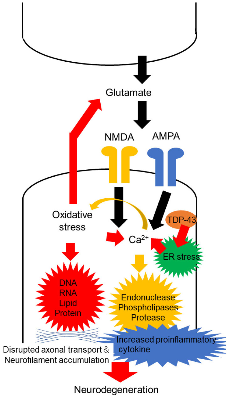
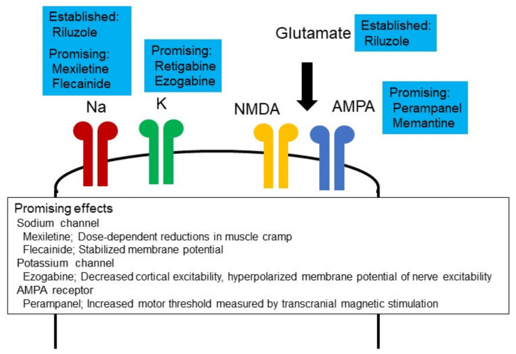
Amyotrophic lateral sclerosis (ALS) is a devastating disease with evidence of degeneration involving upper and lower motor neuron compartments of the nervous system. Presently, two drugs, riluzole and edaravone, have been established as being useful in slowing disease progression in ALS. Riluzole possesses anti-glutamatergic properties, while edaravone eliminates free radicals (FRs). Glutamate is the excitatory neurotransmitter in the brain and spinal cord and binds to several inotropic receptors. Excessive activation of these receptors generates FRs, inducing neurodegeneration via damage to intracellular organelles and upregulation of proinflammatory mediators. FRs bind to intracellular structures, leading to cellular impairment that contributes to neurodegeneration. As such, excitotoxicity and FR toxicities have been considered as key pathophysiological mechanisms that contribute to the cascade of degeneration that envelopes neurons in ALS. Recent advanced technologies, including neurophysiological, imaging, pathological and biochemical techniques, have concurrently identified evidence of increased excitability in ALS. This review focuses on the relationship between FRs and excitotoxicity in motor neuronal degeneration in ALS and introduces concepts linked to increased excitability across both compartments of the human nervous system. Within this cellular framework, future strategies to promote therapeutic development in ALS, from the perspective of neuronal excitability and function, will be critically appraised.

Keywords: amyotrophic lateral sclerosis; cortical excitability; excitotoxicity; free radicals; nerve excitability.


Conflict of interest statement

The authors declare no competing financial interest.

Figures

Figure 1
Figure 1
Neuronal excitability and free radicals accelerate motor neuron degeneration in ALS. Excessive activation of α-amino-3-hydroxy-5-methyl-4-isoxazolepropionate (AMPA) and N-methyl-D-aspartate (NMDA) receptors, mediated by glutamate, increase the influx of calcium ions (Ca2+). Ca2+ activates enzymes such as endonucleases, phospholipases and proteases, which may induce neuronal injury. Additionally, excessive activation of glutamate receptors generates free radicals and oxidative stress to thereby modulate DNA, RNA, lipids and proteins, increase glutamate concentration in the synaptic cleft and raise Ca2+ influx into the cells. These processes accumulate neurofilaments and increase proinflammatory cytokines, which might result in neurodegeneration. Moreover, accumulation of TAR DNA-binding protein 43 (TDP-43) in the cytoplasm of motor neurons potentially induces ER stress, and ER stress leads to the release of Ca2+ from the ER lumen.
Figure 2
Figure 2
Neuronal excitability and therapeutic development. Ion channel modulators have been investigated for efficacy in ALS. Riluzole inhibits glutamate release, enhances glutamate uptake, blocks voltage-dependent sodium channels, antagonizes NMDA receptors and inhibits GABA uptake. Perampanel exerts effects via AMPA receptors, retigabine and ezogabine through potassium channels and mexiletine and flecainide via sodium channels (see text for clinical trial results). These ion channel modulators showed several lines of evidence.

Similar articles

Cited by

References

    1. Mehta P., Raymond J., Punjani R., Larson T., Han M., Bove F., Horton D.K. Incidence of amyotrophic lateral sclerosis in the united states, 2014–2016. Amyotroph. Lateral Scler. Front. Degener. 2022:1–5. doi: 10.1080/21678421.2021.2023190. - DOI - PubMed
    1. Hulisz D. Amyotrophic lateral sclerosis: Disease state overview. Am. J. Manag. Care. 2018;24:S320–S326. - PubMed
    1. Takeda T., Kitagawa K., Arai K. Phenotypic variability and its pathological basis in amyotrophic lateral sclerosis. Neuropathol. Off. J. Jpn. Soc. Neuropathol. 2020;40:40–56. doi: 10.1111/neup.12606. - DOI - PubMed
    1. Giordano A., De Panfilis L., Perin M., Servidio L., Cascioli M., Grasso M.G., Lugaresi A., Pucci E., Veronese S., Solari A. Advance care planning in neurodegenerative disorders: A scoping review. Int. J. Environ. Res. Public Health. 2022;19:803. doi: 10.3390/ijerph19020803. - DOI - PMC - PubMed
    1. Brown C.A., Lally C., Kupelian V., Flanders W.D. Estimated prevalence and incidence of amyotrophic lateral sclerosis and sod1 and c9orf72 genetic variants. Neuroepidemiology. 2021;55:342–353. doi: 10.1159/000516752. - DOI - PubMed