. 2022 Apr 12;15(4):465.
doi: 10.3390/ph15040465.

Effects of a Short-Term Lipopolysaccharides Challenge on Mouse Brain and Liver Peroxisomal Antioxidant and β-oxidative Functions: Protective Action of Argan Oil

Affiliations

Effects of a Short-Term Lipopolysaccharides Challenge on Mouse Brain and Liver Peroxisomal Antioxidant and β-oxidative Functions: Protective Action of Argan Oil

Soukaina Essadek et al. Pharmaceuticals (Basel). .

Abstract

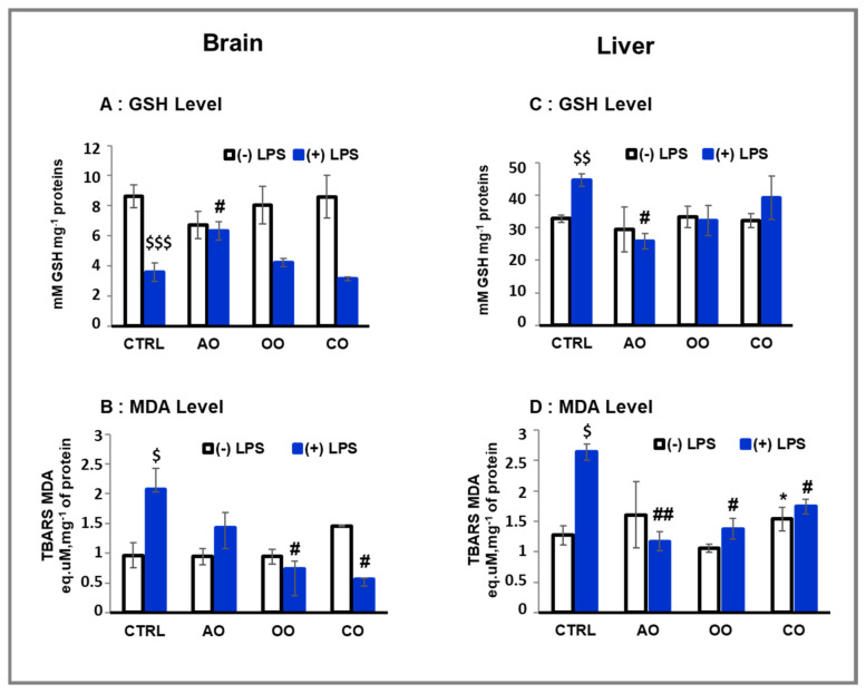
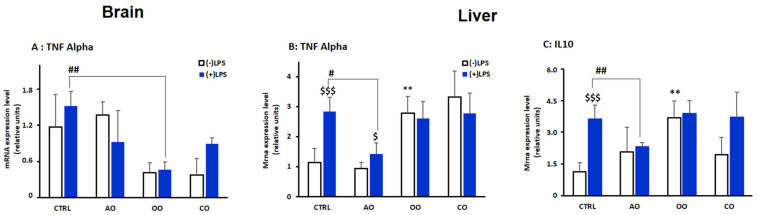
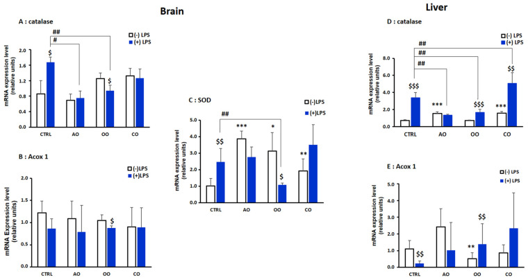
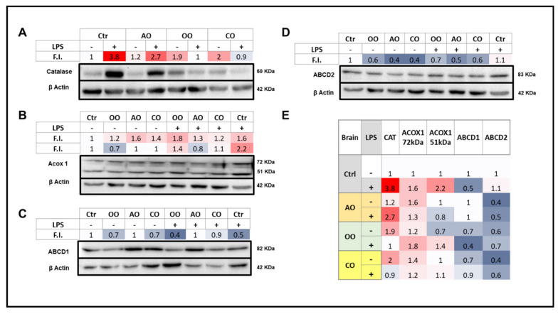
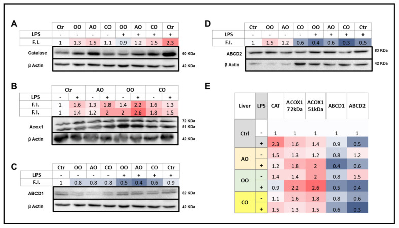
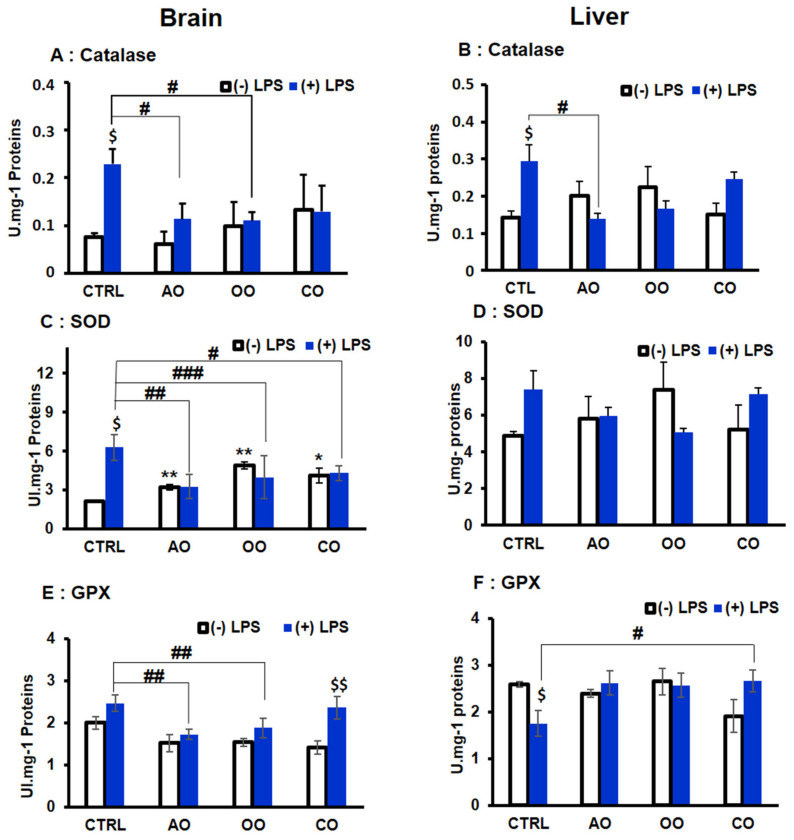
During sepsis, the imbalance between oxidative insult and body antioxidant response causes the dysfunction of organs, including the brain and liver. Exposing mice to bacterial lipopolysaccharides (LPS) results in a similar pathophysiological outcome. The protection offered by argan oil was studied against LPS-induced oxidative stress, dysregulation of peroxisomal antioxidants, and β-oxidation activities in the brain and liver. In a short-term LPS treatment, lipid peroxidation (malonaldehyde assay) increased in the brain and liver with upregulations of proinflammatory tumor necrosis factor (Tnf)-α and anti-inflammatory interleukin (Il)-10 genes, especially in the liver. Although exposure to olive oil (OO), colza oil (CO), and argan oil (AO) prevented LPS-induced lipid peroxidation in the brain and liver, only AO exposure protected against liver inflammation. Remarkably, only exposure to AO prevented LPS-dependent glutathione (GSH) dysregulation in the brain and liver. Furthermore, exposure to AO increased more efficiently than OO and CO in both organs, peroxisomal antioxidant capacity via induction of catalase (Cat) gene, protein and activity expression levels, and superoxide dismutase (Sod1) mRNA and activity levels. Interestingly, LPS decreased protein levels of the peroxisomal fatty acid-ATP binding cassette (ABC) transporters, ABCD1 and ABCD2, and increased acyl-CoA oxidase 1 (ACOX1) protein expression. Moreover, these LPS effects were attenuated for ABCD1 and ACOX1 in the brain of mice pretreated with AO. Our data collectively highlight the protective effects of AO against early oxidative stress caused by LPS in the brain and liver and their reliance on the preservation of peroxisomal functions, including antioxidant and β-oxidation activities, making AO a promising candidate for the prevention and management of sepsis.

Keywords: Acyl-CoA oxidase 1; LPS; antioxidant; argan oil; beta-oxidation; brain; catalase; glutathione peroxidase; peroxisome; superoxide dismutase.


Conflict of interest statement

The authors declare no conflict of interest.

Figures

Figure 1
Figure 1
Effect of argan oil, olive oil, and colza oil treatments on GSH level (A,C) and lipid peroxidation (B,D) in brain and liver. Male C57BL/6 mice received for 28 days a standard diet (control: CTRL), a diet enriched with 6% (w/w) AO, OO, or CO, and an intravenous injection of LPS (100µg) four hours antemortem. All values are means ± SD (n = 3), Statistical significance of higher mean signal strength (* p ≤ 0.05) compared to control, (## p ≤ 0.01, # p ≤ 0.05) compared to LPS and ($$$ p ≤ 0.001, $$ p ≤ 0.01, $ p ≤ 0.05) compared to the different treatments with or without LPS administration.
Figure 2
Figure 2
Effect of argan oil, olive oil, or colza oil treatment on gene expression of the proinflammatory marker Tnf-α (A,B) in the brain and liver, respectively, and on the anti-inflammatory marker Il-10 in the liver (C). Male C57BL/6 mice received for 28 days a standard diet (control (CTRL)), a diet enriched with 6% (w/w) AO, OO, or CO, and intravenous injection of LPS (100 µg) four hours antemortem. First, total RNA was isolated from mice brains or livers, and then the expression level of genes of interest was quantified by real-time RT-qPCR. All values are means ± SD (n = 3), Statistical significance of higher mean signal strength (** p ≤ 0.01) compared to control, (## p ≤ 0.01. # p ≤ 0.05) Compared to LPS and ($$$ p ≤ 0.001. $ p ≤ 0.05) compared to the different treatments with or without LPS administration.
Figure 3
Figure 3
Effect of argan oil, olive oil, or colza oil treatment on gene expression of Cat (A,D) and Acox1 (B,E) in brain and liver, respectively, and Sod in the brain (C). Male C57BL/6 mice received for 28 days a standard diet (control (CTRL)), a diet enriched with 6% (w/w) AO, OO, or CO, and intravenous injection of LPS (100 µg) four hours antemortem. First, total RNA was isolated from mice brains and livers, and then the expression level of genes of interest was quantified by real-time RT-qPCR. All values are means ± SD (n = 3), Statistical significance of higher mean signal strength (*** p ≤ 0.001. ** p ≤ 0.01. * p ≤ 0.05) compared to control, (## p ≤ 0.01. # p ≤ 0.05) Compared to LPS and ($$$ p ≤ 0.001. $$ p ≤ 0.01. $ p ≤ 0.05) compared to the different treatments with or without LPS administration.
Figure 4
Figure 4
Effect of argan oil, olive oil or colza oil treatment on brain expressions of peroxisomal proteins, CAT (A), ACOX1 (B), ABCD1 (C), ABCD2 (D) and the heatmap for all protein expression (E). Male C57BL/6 mice received for 28 days a standard diet (control (CTRL)), a diet enriched with 6% (w/w) AO, OO, or CO, and intravenous injection of LPS (100 µg) four hours antemortem. Brain homogenates were separated in PAGE-SDS electrophoresis and subjected to immunoblotting as described in Section 4. Band intensities were analyzed by densitometry and standardized to β-actine expression level. Tables represent the standardized densitometric analysis obtained after signal intensity quantification of different proteins.
Figure 5
Figure 5
Effect of argan oil, olive oil or colza oil treatment on liver expressions of peroxisomal proteins, CAT (A), ACOX1 (B), ABCD1 (C), ABCD2 (D) and the heatmap for all protein expression (E). Male C57BL/6 mice received for 28 days a standard diet (control (CTRL)), a diet enriched with 6% (w/w) AO, OO, or CO, and intravenous injection of LPS (100 µg) four hours antemortem. Liver homogenates were prepared as described in Section 4. Band intensities were analyzed by densitometry and standardized to β-actin expression level. Tables represent the standardized densitometric analysis obtained after signal intensity quantification of different proteins.
Figure 6
Figure 6
Effect of argan oil, olive oil or colza oil treatment on the antioxidant enzymes activities of CAT (A,B), SOD (C,D), and GPX (E,F) in brain and liver, respectively. C57BL/6 mice received for 28 days a standard diet (control (CTRL)), a diet enriched with 6% (w/w) AO, OO, or CO, and intravenous injection of LPS (100 µg) four hours antemortem. Brain and liver homogenates were prepared as described in Section 4. Results are expressed in (UI.mg−1 = µmol of substrate transformed/minute/mg of proteins). All values are means ± SD (n = 3), statistical significance of higher mean signal strength (** p ≤ 0.01. * p ≤ 0.05) compared to control, (### p ≤ 0.001, ## p ≤ 0.01, # p ≤ 0.05), compared to LPS and $$ p ≤ 0.01, $ p ≤ 0.05) compared to treatment with or without LPS administration.

References

    1. Sharshar T., Annane D., de la Grandmaison G.L., Brouland J.P., Hopkinson N.S., Françoise G. The Neuropathology of Septic Shock. Brain Pathol. Zurich Switz. 2004;14:21–33. doi: 10.1111/j.1750-3639.2004.tb00494.x. - DOI - PMC - PubMed
    1. Mantzarlis K., Tsolaki V., Zakynthinos E. Role of Oxidative Stress and Mitochondrial Dysfunction in Sepsis and Potential Therapies. Oxid. Med. Cell. Longev. 2017;2017:5985209. doi: 10.1155/2017/5985209. - DOI - PMC - PubMed
    1. Sriskandan S., Altmann D.M. The Immunology of Sepsis. J. Pathol. 2008;214:211–223. doi: 10.1002/path.2274. - DOI - PubMed
    1. Spolarics Z. Endotoxemia, Pentose Cycle, and the Oxidant/Antioxidant Balance in the Hepatic Sinusoid. J. Leukoc. Biol. 1998;63:534–541. doi: 10.1002/jlb.63.5.534. - DOI - PubMed
    1. Fransen M., Lismont C. Peroxisomes and Cellular Oxidant/Antioxidant Balance: Protein Redox Modifications and Impact on Inter-Organelle Communication. Subcell. Biochem. 2018;89:435–461. doi: 10.1007/978-981-13-2233-4_19. - DOI - PubMed

LinkOut - more resources