. 2022 Apr 18;15(4):490.
doi: 10.3390/ph15040490.

Development of Astaxanthin-Loaded Nanosized Liposomal Formulation to Improve Bone Health

Affiliations

Development of Astaxanthin-Loaded Nanosized Liposomal Formulation to Improve Bone Health

Hsin-I Chang et al. Pharmaceuticals (Basel). .

Abstract

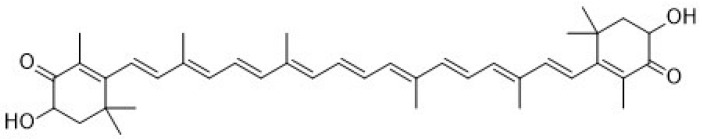
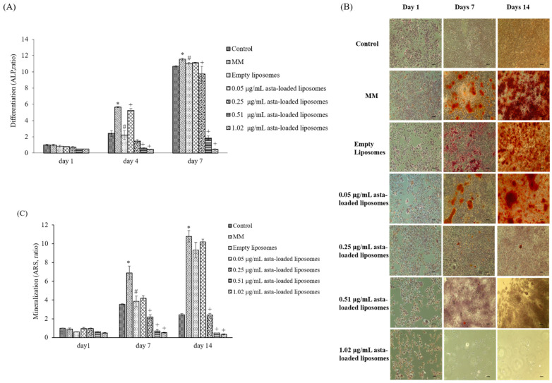
Astaxanthin is a xanthophyll carotenoid commonly found in marine organisms. Due to its super antioxidative ability, astaxanthin has been widely applied as a human nutraceutical supplement for health benefits. In order to enhance the bioavailability of astaxanthin, we used soybean phosphatidylcholine to encapsulate astaxanthin for liposomal formation. The physical properties of astaxanthin (asta)-loaded liposomes were determined by particle size, encapsulation efficiency and polydispersity index. The results revealed that the particle sizes of asta-loaded liposomes with various concentrations exhibited mean diameters in the range of 109 to 134 nm and had a narrow PDI value. As expected, the entrapment efficiency of liposomes loaded with a low concentration of astaxanthin (0.05 μg/mL) was 89%, and that was reduced to 29% for 1.02 μg/mL asta loading. Alizarin red staining and calcium content measurement showed that there was a significant reduction in calcium deposition for 7F2 osteoblasts treated with asta-loaded liposomes (0.25-1.02 μg/mL) in comparison with the cells treated with drug-free liposomes and mineralization medium (MM). Although liposomal formulation can reduce the cytotoxicity of astaxanthin and possess antioxidant, anti-inflammatory and anti-osteoclastogenic activities in RAW264.7 macrophages, asta-loaded liposomes with high concentrations may suppress ALP activity and mineralization level in 7F2 osteoblasts. Therefore, astaxanthin extract may be able to protect bones against oxidative stress and inflammation through liposomal formulation.

Keywords: anti-inflammation; astaxanthin; liposomes; marine natural product; osteoblast mineralization.


Conflict of interest statement

The authors declare no conflict of interest.

Figures

Figure 1
Figure 1
The chemical structure of astaxanthin.
Figure 2
Figure 2
The antioxidant ability of astaxanthin extract measured by DPPH (A) and (B) ABTS assays.
Figure 3
Figure 3
Physical stabilities of liposomal formulations. (A) Asta-loaded and empty liposomes were stored at 4 °C in DMEM containing 10% FBS over 1, 4, 7 and 14 days of incubation. (B) Asta-loaded and empty liposomes were stored at 37 °C in DMEM containing 10% FBS and incubated for 1, 4, 7 and 14 days. The particle sizes are presented as the mean ± standard deviation.
Figure 4
Figure 4
In vitro uptake of DiI-labeled liposomes in 7F2 osteoblasts measured by fluorescent microscopy. The cells were cultured with DiI-labeled liposomes for 4 h. Thereafter, red fluorescence (DiI-labeled liposomes) and blue fluorescence (DAPI for nuclei staining) were evaluated by fluorescent microscopy equipped with a CCD system (×100 magnification, scale bar = 200 nm).
Figure 5
Figure 5
The effect of astaxanthin extract and asta-loaded liposomes on cell viability of Raw264.7 mouse macrophages. Macrophages were treated with (A) astaxanthin extract and (B) empty and asta-loaded liposomes for 24 h. Viabilities of Raw264.7 macrophages were measured by MTT assay. The data are shown as the means ± standard deviation. (* p < 0.05 related to control; + p < 0.05 related to 2% ETOH; # p < 0.05 related to liposomes).
Figure 6
Figure 6
The effect of astaxanthin extract and asta-loaded liposomes on cell viability of 7F2 osteoblasts. 7F2 osteoblasts were treated with (A) astaxanthin extract and (B) empty and asta-loaded liposomes for 24 h. Cell viabilities of 7F2 osteoblasts were measured by MTT assay. The data are shown as the means ± standard deviation. (* p < 0.05 related to control; + p < 0.05 related to 2% ETOH; # p < 0.05 related to liposomes).
Figure 7
Figure 7
The effects of asta-loaded liposomes on nitrite production, Cox-2 expression and TRAP activity of LPS-induced Raw264.7 mouse macrophages. Raw264.7 macrophages were co-treated with LPS and asta-loaded liposomes overnight. (A) NO production in no-phenol red medium was evaluated by chemical Griess reagent. Nitric oxide assay is a colorimetric method, once the Griess reagents contact the solution containing nitrite ions, the reaction mixture will change color to pink; the absorbance was measured at OD 550 nm. The nitrite production in LPS-induced cells is presented as 100% (* p < 0.05 related to control, + p < 0.05 related to LPS and # p < 0.05 related to cells treated with empty liposomes). (B) COX-2 mRNA expression of Raw264.7 macrophages. Real-time PCR analysis was performed in Raw264.7 macrophages after 24 h treatment with LPS (* p < 0.05 related to control and # p < 0.05 related to LPS). (C) Asta-loaded liposomes inhibit TRAP activity in LPS- and RANKL-induced RAW264.7 macrophages. Data are expressed as the percentage of TRAP activity measured in cells derived from each model in comparison with control (* p < 0.05 related to control and # p < 0.05 related to cells treated with LPS and RANKL).
Figure 8
Figure 8
The effect of asta-loaded liposomes on intracellular ROS production of LPS-induced Raw264.7 mouse macrophages. Raw264.7 macrophages were co-treated with LPS and asta-loaded liposomes overnight. (A) Production of reactive oxygen species (ROS) (×100 magnification, scale bar = 200 nm). (B) Quantification of relative fluorescence intensity. Results are revealed as percentages with mean ± standard deviation (n = 4–6). Fluorescence images and quantitation of relative fluorescence intensity representing cytosolic ROS detection by DCF-DA staining (* p < 0.05 related to control, + p < 0.05 related to LPS and # p < 0.05 related to cells treated with empty liposomes).
Figure 9
Figure 9
The effect of asta-loaded liposomes on 7F2 osteoblast differentiation and mineralization. 7F2 osteoblasts were treated with 50 μg/mL of ascorbic and 10 mM β-glycerophosphate to induce osteoblast differentiation and mineralization. (A) Cells were then incubated in the presence or absence of asta-loaded liposomes for 1, 4 and 7 days. Results are revealed as a ratio with mean ± standard deviation (n = 3). (B) ARS staining of calcium deposits on days 1, 7 and 14 (×100 magnification, scale bar = 200 nm). (C) Quantification of calcium deposits on days 1, 7 and 14. Calcium deposition was quantified by dissolving ARS aggregates into 10% cetylpyridinium chloride and evaluating the absorbance at 560 nm. Results are revealed as a ratio with mean ± standard deviation (n = 3) (* p < 0.05 related to control group of the same day, # p < 0.05 related to MM of the same day, + p < 0.05 related to empty liposome treatment of the same day).

Similar articles

Cited by

References

    1. Ambati R.R., Phang S.M., Ravi S., Aswathanarayana R.G. Astaxanthin: Sources, extraction, stability, biological activities and its commercial applications—A review. Mar. Drugs. 2014;12:128–152. doi: 10.3390/md12010128. - DOI - PMC - PubMed
    1. Yamashita E. Astaxanthin as a Medical Food. J. Funct. Foods Health Dis. 2013;3:254–258. doi: 10.31989/ffhd.v3i7.49. - DOI
    1. Miki W. Biological Functions and Activities of Animal Carotenoids. Pure Appl. Chem. 1991;63:141–146. doi: 10.1351/pac199163010141. - DOI
    1. Terao J. Antioxidant activity of β-carotene-related carotenoids in solution. Lipids. 1989;24:659–661. doi: 10.1007/BF02535085. - DOI - PubMed
    1. Jyonouchi H., Sun S., Iijima K., Gross M.D. Antitumor activity of astaxanthin and its mode of action. Nutr. Cancer. 2000;36:59–65. doi: 10.1207/S15327914NC3601_9. - DOI - PubMed