Review
. 2022 Apr 15;9(4):565.
doi: 10.3390/children9040565.

'Teratoid' Hepatoblastoma: An Intriguing Variant of Mixed Epithelial-Mesenchymal Hepatoblastoma

Affiliations
Review

'Teratoid' Hepatoblastoma: An Intriguing Variant of Mixed Epithelial-Mesenchymal Hepatoblastoma

Consolato M Sergi et al. Children (Basel). .

Abstract

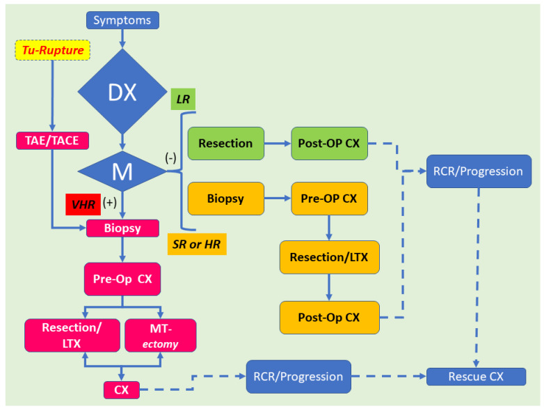
Liver neoplasms are quite rare in childhood. They often involve 6.7 cases per 10 million children aged 18 years or younger. Hepatoblastoma (HB) is the most frequent tumor, but this neoplasm's rarity points essentially to the difficulty of performing biologic studies and large-scale therapeutic trials. On the pathological ground, HB is separated into an entirely epithelial neoplasm or a mixed neoplasm with epithelial and mesenchymal components. This last category has been further subdivided into harboring teratoid features or not. The 'teratoid' HB includes a mixture of components with heterologous origin. The heterologous components include neuroectoderm, endoderm, or melanin-holding cells with or without mesenchymal components. The most important criterium for the teratoid component is neuroepithelium, melanin, and, more recently, a yolk-sac-like component and neuroendocrine components. The mesenchymal components include muscle, osteoid, and cartilage, which are most often observed mainly in 'teratoid' neoplasms. The teratoid component or mesenchymal components are diagnosed with biopsies. They appear more prominent after chemotherapy due to the response and shrinkage of epithelial elements and non- or low-responsive components of mixed HB. This review focuses on the clinical, radiological, and pathological findings of HB with teratoid features.

Keywords: cancer; liver; management; radiology.


Conflict of interest statement

The authors declare no conflict of interest.

Figures

Figure 1
Figure 1
The schema depicts the most current classification of the subgroups of hepatoblastoma. The teratoid hepatoblastoma or hepatoblastoma with teratoid features is part of the mixed type (epithelial and mesenchymal components) of hepatoblastoma.
Figure 2
Figure 2
Imaging of a teratoid hepatoblastoma showing a heterogeneous mass encased in the liver (a), which is very close to the diaphragm (b). The magnetic resonance imaging (cg) shows a large neoplasm in the right upper quadrant of the abdomen deriving from the right liver lobe. The neoplasm has two main portions and seems to contain different portions of tissue. The coronal view showed that the tumor is exophytic, protruding superiorly (T2 sequence). Remarkably, the transversal view typically reveals that the ventral tumor portion has a cystic nonenhancing appearance, whereas the dorsal part emerges more solid (T2 sequence). The T1 sequences—after contrast (post-gadolinium) application—(eg) display an inhomogeneous contrast enhancement. The ventral portion of the hepatic neoplasm is intriguingly avidly enhancing, while the dorsal solid portion enhances less avidly.
Figure 3
Figure 3
Histology of a mixed hepatoblastoma with teratoid features or teratoid hepatoblastoma showing fetal and embryonal portions and teratoid features with a yolk-sac-like pattern. Cords and solid portions are present and numerous mitoses are recognized with epithelial and mesenchymal components and yolk-sac-like elements (d), ((ad) hematoxylin and eosin staining, X50 original magnification, scale bar, 100 microns).
Figure 4
Figure 4
Immunohistochemistry of a teratoid hepatoblastoma. The microphotographs show positivity for polyclonal carcino-embryonic antigen (pCEA) ((a), Avidin-Biotin Complex anti-pCEA immunostaining, ×100 original magnification, scale bar, 50 microns), β-catenin ((b), Avidin-Biotin Complex anti-β-catenin immunostaining, ×100 original magnification, scale bar, 50 microns), (cyto-)keratin 19 (CK19) ((c), Avidin-Biotin Complex anti-K19 immunostaining, ×100 original magnification, scale bar, 50 microns), and α-fetoprotein (AFP) ((d), Avidin-Biotin Complex anti-AFP immunostaining, ×100 original magnification, scale bar, 50 microns).
Figure 5
Figure 5
Diagnosis and therapeutic algorithm for hepatoblastoma. Hepatoblastoma is typically diagnosed by objective clinical signs, imaging data, and an increase in serum α-fetoprotein (AFP) levels. In some children affected with hepatoblastoma harboring a tumor, which is identified as ruptured, transarterial embolization (TAE) or transarterial chemoembolization (TACE) is performed to monitor the intraperitoneal hemorrhage. After the hemorrhage is under control, these children should be handled according to the very high-risk stratification flow. Amongst the children without distant tumor spreading [M(−)], low-risk (LR) children are approached with primary resection followed by chemotherapy in the postoperative period (Post-op CX). Standard-risk (SR) (also known as “intermediate-risk” (IR)) or high-risk (HR) children with HB receive chemotherapy in the preoperative period (Pre-op CX). Then, these patients undergo primary tumor resection by hepatectomy or liver transplantation (LTX). The very high-risk (VHR) patients affected by HB and distant tumor spreading (M(+))receive Pre-op CX. Then, a patient whose distant tumor spreading is reduced by CX goes through primary tumor resection by hepatectomy. Alternatively, these patients may undergo LTX, followed by Post-op CX. However, a patient whose distant tumor spreading remains needs to go through “metastasectomy” or hepatectomy. LTX is typically generally for the patient devoid of distant metastasis. On the other side, LTX may be indicated when distant tumor spreading has obviously vanished. Finally, patients with recurrence (RCR) or neoplastic progression should undertake some kind of rescue chemotherapy, also known as salvage CX, which is a form of therapy given after a disease does not react to standard therapy.

Similar articles

Cited by

References

    1. Sharma D., Subbarao G., Saxena R. Hepatoblastoma. Semin. Diagn. Pathol. 2017;34:192–200. doi: 10.1053/j.semdp.2016.12.015. - DOI - PubMed
    1. Saeed O., Saxena R. Primary mesenchymal liver tumors of childhood. Semin. Diagn. Pathol. 2017;34:201–207. doi: 10.1053/j.semdp.2016.12.016. - DOI - PubMed
    1. Kromhout H., Friesen M., Marques M.M., Sergi C.M., Abdallah M., Benke G., Cesta M., Germolec D., Houck K., Ichihara G., et al. Carcinogenicity of isobutyl nitrite, beta-picoline, and some acrylates. Lancet Oncol. 2018;19:1020–1022. doi: 10.1016/S1470-2045(18)30491-1. - DOI - PubMed
    1. Guyton K.Z., Loomis D., Grosse Y., El Ghissassi F., Benbrahim-Tallaa L., Guha N., Scoccianti C., Mattock H., Straif K., International Agency for Research on Cancer Monograph Working Group Carcinogenicity of tetrachlorvinphos, parathion, malathion, diazinon, and glyphosate. Lancet Oncol. 2015;16:490–491. doi: 10.1016/S1470-2045(15)70134-8. - DOI - PubMed
    1. Guyton K.Z., Loomis D., Grosse Y., El Ghissassi F., Bouvard V., Benbrahim-Tallaa L., Guha N., Mattock H., Straif K., International Agency for Research on Cancer Monograph Working Group Carcinogenicity of pentachlorophenol and some related compounds. Lancet Oncol. 2016;17:1637–1638. doi: 10.1016/S1470-2045(16)30513-7. - DOI - PubMed

LinkOut - more resources