

Cranioplasty after Two Giant Intraosseous Angiolipomas of the Cranium: Case Report and Literature Review
- PMID: 35455833
- PMCID: PMC9028485
- DOI: 10.3390/healthcare10040655
Cranioplasty after Two Giant Intraosseous Angiolipomas of the Cranium: Case Report and Literature Review
Abstract
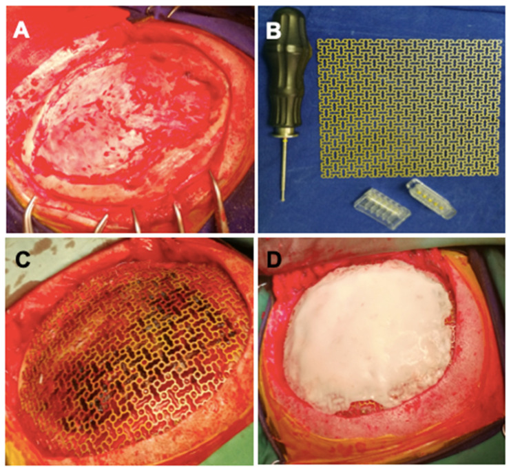
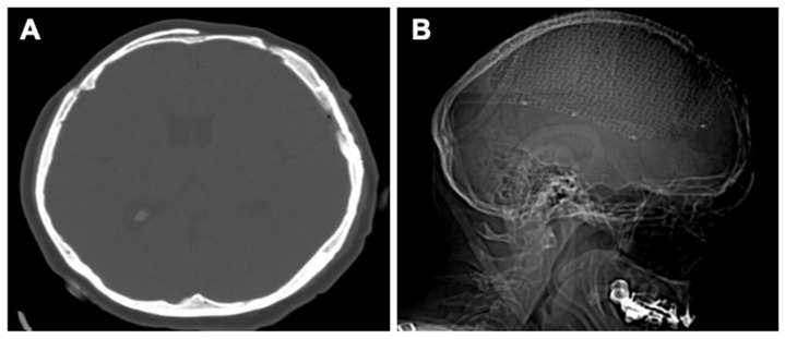
Angiolipomas are rare, benign tumors resulting from the proliferation of adipose tissue and blood vessels, most frequently encountered subcutaneously at the upper limbs and trunk level. Due to their rarity, few cases of intraosseous angiolipomas are presented in the literature. The paper reports a 50-year-old female case with intracranial hypertension syndrome, frontal and parietal headache, nausea, and vomiting symptoms increasing in intensity. A CT exam revealed two hypodense expansive intraosseous formations/lesions. The first one was located in the projection of the frontal bone and the second one was placed on the left parietal bone. After further investigations, a two-stage procedure was considered. A frontal craniotomy with excision of the intraosseous tumor was performed in the first stage. In the second stage, a left parietal craniotomy was done with excision of the intraosseous tumor combined with a cranioplasty procedure. The patient had a favorable postoperative evolution with no symptoms or neurological deficits. This is among the few reported cases of intraosseous angiolipoma located at the cranium level and the first case report of two intraosseous angiolipomas situated on the same site. The medical recommendation was a complete surgical excision of the lesion followed by cranioplasty.
Keywords: bone tumor; cranioplasty; craniotomy; cranium; intraosseous angiolipoma; rare disease; two stage surgical procedure.
Conflict of interest statement
The authors declare no conflict of interest.
Figures


References
-
- Moldovan H., Popescu D., Buliga T., Filip A., Antoniac I., Gheorghiţă D., Molnar A. Gastric Adenocarcinoma Associated with Acute Endocarditis of the Aortic Valve and Coronary Artery Disease in a 61-Year-Old Male with Multiple Comorbidities—Combined Surgical Management—Case Report. Medicina. 2019;55:242. doi: 10.3390/medicina55060242. - DOI - PMC - PubMed
-
- Dumitrescu D., Savlovschi C., Borcan R., Pantu H., Serban D., Gradinaru S., Smarandache G., Trotea T., Branescu C., Musat L., et al. Caz Clinic—Hernie Diafragmatica Voluminoasa—Abdomen Acut Chirurgical: Dificultati Diagnostice Si Terapeutice. Chirurgia. 2011;106:657–660. - PubMed
-
- Lee C.H., Son D.W., Lee S.H., Lee J.S., Lee S.W., Song G.S. Spinal Epidural Angiolipoma: A Case Report on Sudden Hemorrhagic Onset and Review of the Literature. J. Korean Soc. Geriatr. Neurosurg. 2021;17:20–24. doi: 10.51638/jksgn.2021.00038. - DOI
-
- Fernando H., Mohammad S., Baarini O., Haddad A. Angiolipoma of the First Intermetatarsal Space—A Case Study. Foot Ankle Surg. Tech. Rep. Cases. 2022;2:100177. doi: 10.1016/j.fastrc.2022.100177. - DOI
LinkOut - more resources
Full Text Sources
