Review
. 2022 Apr 12;11(8):1312.
doi: 10.3390/cells11081312.

Advanced Glycation End-Products (AGEs): Formation, Chemistry, Classification, Receptors, and Diseases Related to AGEs

Affiliations
Review

Advanced Glycation End-Products (AGEs): Formation, Chemistry, Classification, Receptors, and Diseases Related to AGEs

Aleksandra Twarda-Clapa et al. Cells. .

Abstract

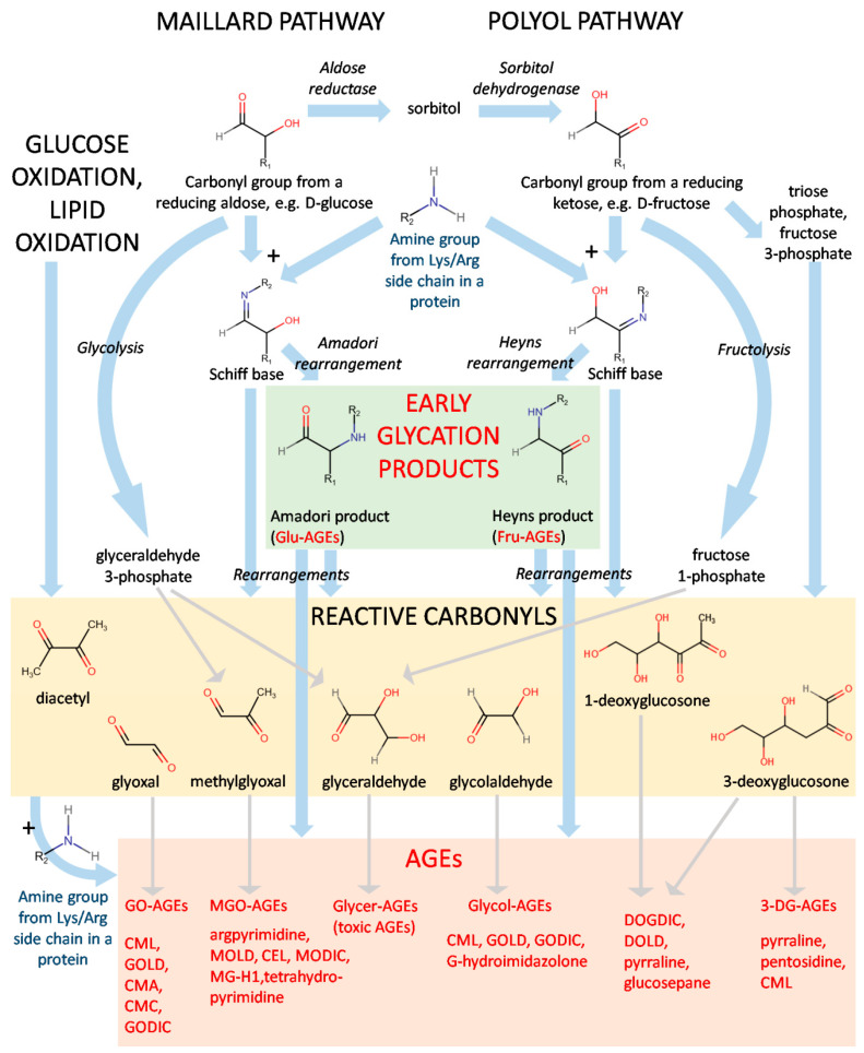
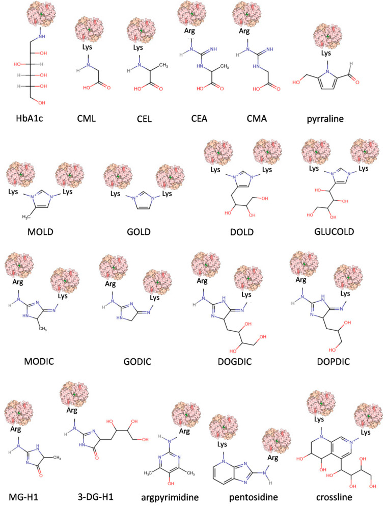
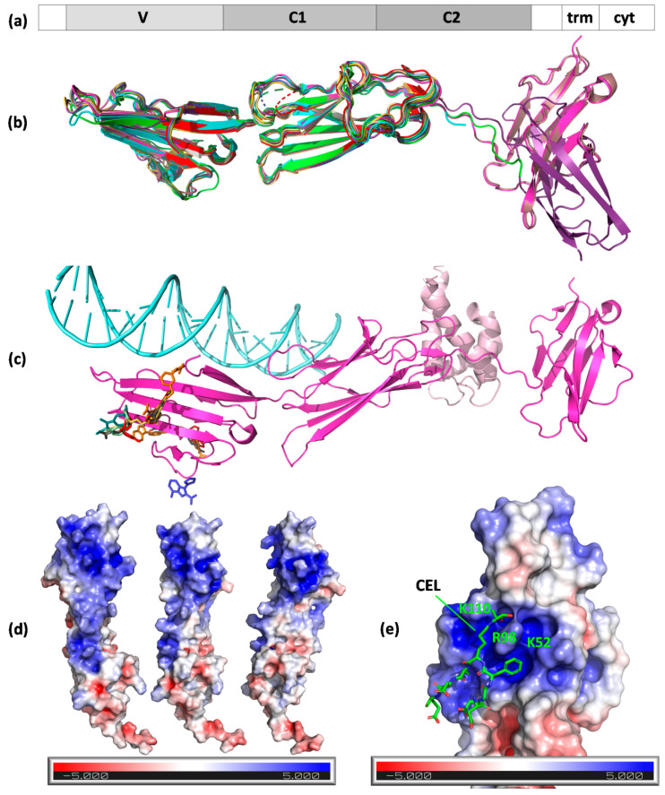
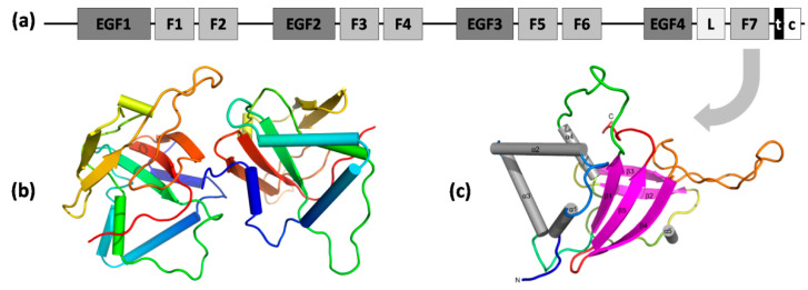
Advanced glycation end-products (AGEs) constitute a non-homogenous, chemically diverse group of compounds formed either exogeneously or endogeneously on the course of various pathways in the human body. In general, they are formed non-enzymatically by condensation between carbonyl groups of reducing sugars and free amine groups of nucleic acids, proteins, or lipids, followed by further rearrangements yielding stable, irreversible end-products. In the last decades, AGEs have aroused the interest of the scientific community due to the increasing evidence of their involvement in many pathophysiological processes and diseases, such as diabetes, cancer, cardiovascular, neurodegenerative diseases, and even infection with the SARS-CoV-2 virus. They are recognized by several cellular receptors and trigger many signaling pathways related to inflammation and oxidative stress. Despite many experimental research outcomes published recently, the complexity of their engagement in human physiology and pathophysiological states requires further elucidation. This review focuses on the receptors of AGEs, especially on the structural aspects of receptor-ligand interaction, and the diseases in which AGEs are involved. It also aims to present AGE classification in subgroups and to describe the basic processes leading to both exogeneous and endogeneous AGE formation.

Keywords: AGE classification; AGE formation; AGE receptors; AGE-related diseases; AGEs; RAGE; Stab2; advanced glycation end-products.


Conflict of interest statement

The authors declare no conflict of interest. The funders had no role in the design of the study; in the collection, analyses, or interpretation of data; in the writing of the manuscript; or in the decision to publish the results.

Figures

Figure 1
Figure 1
A scheme of formation for AGEs. Abbreviations: glucose-derived (Glu-AGEs), fructose-derived (Fru-AGEs), glyoxal-derived (GO-AGEs), methylglyoxal-derived (MGO-AGEs), glyceraldehyde-derived (Glycer-AGEs), glycolaldehyde-derived (Glycol-AGEs), and 3-deoxyglucosone-derived (3-DG-AGEs).
Figure 2
Figure 2
Chemical structure representations of AGEs. Abbreviations: glycated hemoglobin (HbA1c), Nε–(carboxymethyl)lysine (CML), Nε-(1-carboxyethyl)lysine (CEL), N7–(1-carboxyethyl)arginine (CEA), N7–(carboxymethyl)arginine (CMA), 6-(2-formyl-5-hydroxymethyl-1-pyrrolyl)-L-norleucine (pyrraline), 6-{1-[(5S)-5-ammonio-6-oxido-6-oxohexyl]-4-methyl-imidazolium-3-yl}-L-norleucine (MOLD), 6-{1-[(5S)-5-ammonio-6-oxido-6-oxohexyl]imidazolium-3-yl}-L-norleucine (GOLD),1,3-di(Nε-lysino)-4-(2,3,4-trihydroxybutyl)-imidazolium (DOLD),1,3-bis-(5-amino-5-carboxypentyl)-4-(1,2,3,4-tetrahydroxybutyl)-3H-imidazolium (GLUCOLD), 2-ammonio-6-({2-[4-ammonio-5-oxido-5-oxopently)amino]4-methyl-4,5-dihydro-1H-imidazol-5-ylidene}amino)hexanoate (MODIC), N6-(2-{4S(-4-ammonio-5-oxido-5-oxopentyl]amino}-3,5-dihydro-4H-imidazol-4-ylidene)-L-lysine (GODIC), N6-{2-{[(4S)-4-ammonio-5-oxido-5-oxopentyl]amino}-5-[(2S,3R)-2,3,4-trihydroxybutyl]-3,5-dihydro-4H-imidazol-4-ylidene}-L-lysinate (DOGDIC), N6-{2-{[(4S)-4-ammonio-5-oxido-5-oxopentyl]amino}-5-[(2S)-2,3-dixydroxypropyl]3,5-dihydro-4H-imidazol-4-ylidene}-L-lysinate (DOPDIC), Nδ-(5-methyl-4-imidazolon-2-yl)-L-ornithine (MG-H1), Nδ-(45-hydro-5-(2,3,4-trihydroxybutyl)-4-imidazolon-2-yl]-L-ornithine (3DG-H1), Nδ-(5-hydroxy-4,6-dimethylpyrimidine-2-yl)-L-ornithine (argpyrimidine), and 6-[2-[[(4S)-4-amino-5-hydroxy-5-oxopentyl]amino]-4-imidazo[4,5-b]pyridinyl]-L-norleucine (pentosidine). Modified protein surface models (light pink) are based on the structure of human hemoglobin (PDB ID 1COH).
Figure 3
Figure 3
Structures of RAGE and ligand binding mode. (a) The domain organization of the receptor: 1–21, signal peptide; 23–116, Ig-like V-type; 124–221, Ig-like C2-type 1; 227–317, Ig-like C2-type 2; 343–363, transmembrane helical; 364–404, cytoplasmic (disordered); (b) A chains (N chain in the case of 3o3u, gold) of RAGE structure models aligned to 3cjj A chains (VC1 fragment, green). Protein chains are shown as cartoons and are colored: 4oi7, cyan; 4lp5, dark violet; 4p2y, dirty pink; 4ybh, pink; 6xq1, yellow; 6xq3, orange; 6xq5, olive; 6xq6, pale green; 6xq7, dark cyan; 6xq8, red; 6xq9, gray; and 7lmw, dark blue. MBP fusion protein in 3o3u is not shown. A chains (N in the case of 3o3u) of RAGE aligned to 3cjj A chain (VC1 fragment) with low or very low RMSD (given in Å): 3o3u–0.991, 4lp5–0.889, 4oi7–0.759, 4p2y–0.544, 4ybh–0.602, 6xq1–1.094, 6xq3–1.221, 6xq5–1.122, 6xq6–1.107, 6xq7–1.161, 6xq8–1.325, 6xq9–1.241, and 7lmw–1.264; (c) RAGE VC1C2 (cartoon, PDB ID 4p2y, magenta) with its ligands (S100-A6, cartoon, light pink, 4p2y; DNA, light cyan, 4oi7) and inhibitors (colored sticks, from PDB IDs 6xq–6xq9 and 7lmw); (d) positively charged patch on the surface of RAGE (surface view, PDB ID 3cjj) shown in three orientations; (e) CEL peptide binding to the positively charged patch on RAGE surface (PDB ID 2l7u).
Figure 4
Figure 4
Domain organization of Stab1 and Stab2 receptors and the structure of FAS1 domain of Stab2. (a) Domains of stabilin receptors: EGF1–4—EGF-like domain repeats (in four series); F1–7—seven FAS1 domains; L—Link domain; t—transmembrane region; c—cytoplasmic (disordered) domain; (b) structure of the seventh FAS1 domain of Stab2 (PDB ID 5N86)—cartoon representation of chains A and B in an asymmetric unit (rainbow from blue N-terminus to red C-terminus); (c) different orientation of chain A with depicted secondary structure elements shown as on the cartoon model.

References

    1. Maillard L.-C. Action Des Acides Aminés Sur Les Sucres: Formation Des Melanoidines Par Voie Méthodique. CR Acad. Sci. 1912;154:66–68.
    1. Rahbar S., Blumenfeld O., Ranney H.M. Studies of an Unusual Hemoglobin in Patients with Diabetes Mellitus. Biochem. Biophys. Res. Commun. 1969;36:838–843. doi: 10.1016/0006-291X(69)90685-8. - DOI - PubMed
    1. Cerami A., Vlassara H., Brownlee M. Role of Nonenzymatic Glycosylation in Atherogenesis. J. Cell. Biochem. 1986;30:111–120. doi: 10.1002/jcb.240300203. - DOI - PubMed
    1. Shen C.Y., Lu C.H., Wu C.H., Li K.J., Kuo Y.M., Hsieh S.C., Yu C.L. The Development of Maillard Reaction, and Advanced Glycation End Product (Age)-Receptor for Age (Rage) Signaling Inhibitors as Novel Therapeutic Strategies for Patients with Age-Related Diseases. Molecules. 2020;25:5591. doi: 10.3390/molecules25235591. - DOI - PMC - PubMed
    1. Chaudhuri J., Bains Y., Guha S., Kahn A., Hall D., Bose N., Gugliucci A., Kapahi P. The Role of Advanced Glycation End Products in Aging and Metabolic Diseases: Bridging Association and Causality. Cell Metab. 2018;28:337–352. doi: 10.1016/j.cmet.2018.08.014. - DOI - PMC - PubMed

Publication types

Substances