. 2022 Apr 13;11(8):1314.
doi: 10.3390/cells11081314.

Transcription Factor CREB3L1 Regulates the Expression of the Sodium/Iodide Symporter (NIS) in Rat Thyroid Follicular Cells

Affiliations

Transcription Factor CREB3L1 Regulates the Expression of the Sodium/Iodide Symporter (NIS) in Rat Thyroid Follicular Cells

Pablo Di Giusto et al. Cells. .

Abstract

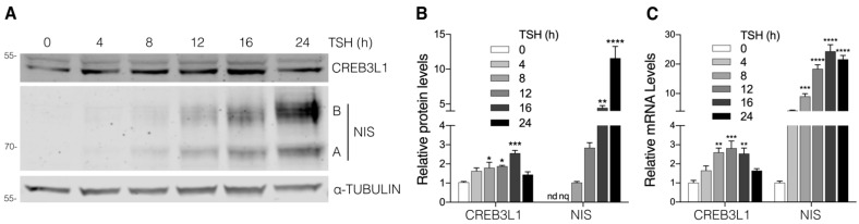
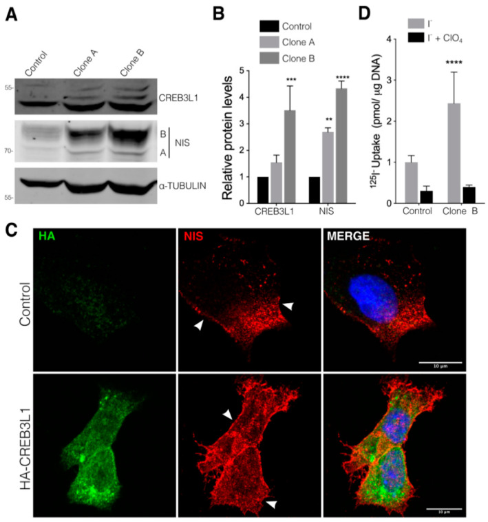
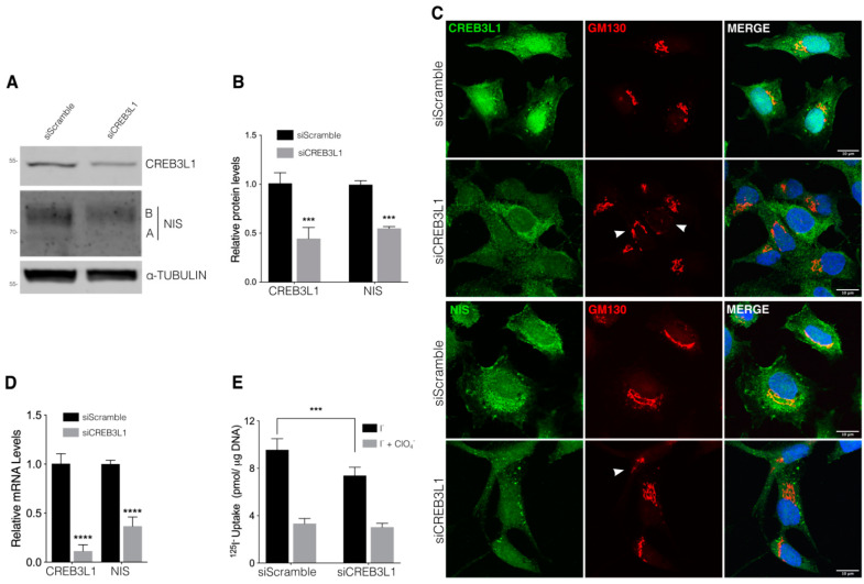
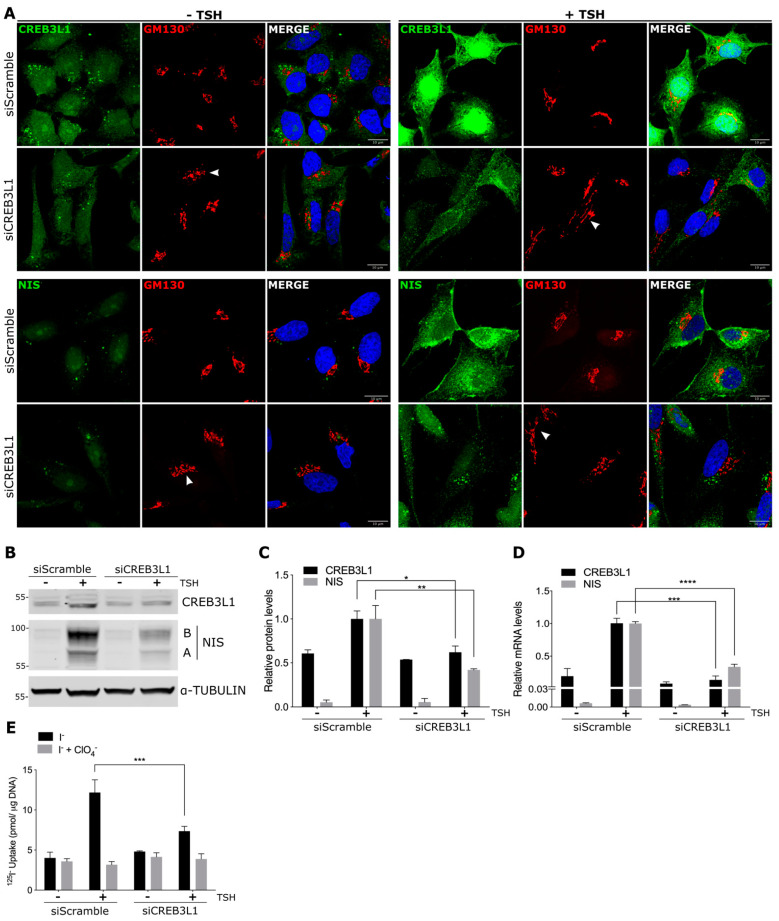
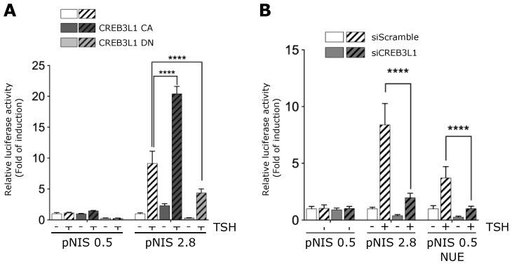
The transcription factor CREB3L1 is expressed in a wide variety of tissues including cartilage, pancreas, and bone. It is located in the endoplasmic reticulum and upon stimulation is transported to the Golgi where is proteolytically cleaved. Then, the N-terminal domain translocates to the nucleus to activate gene expression. In thyroid follicular cells, CREB3L1 is a downstream effector of thyrotropin (TSH), promoting the expression of proteins of the secretory pathway along with an expansion of the Golgi volume. Here, we analyzed the role of CREB3L1 as a TSH-dependent transcriptional regulator of the expression of the sodium/iodide symporter (NIS), a major thyroid protein that mediates iodide uptake. We show that overexpression and inhibition of CREB3L1 induce an increase and decrease in the NIS protein and mRNA levels, respectively. This, in turn, impacts on NIS-mediated iodide uptake. Furthermore, CREB3L1 knockdown hampers the increase the TSH-induced NIS expression levels. Finally, the ability of CREB3L1 to regulate the promoter activity of the NIS-coding gene (Slc5a5) was confirmed. Taken together, our findings highlight the role of CREB3L1 in maintaining the homeostasis of thyroid follicular cells, regulating the adaptation of the secretory pathway as well as the synthesis of thyroid-specific proteins in response to TSH stimulation.

Keywords: CREB3L1; cellular homeostasis; endoplasmic reticulum; iodide uptake; sodium/iodide symporter (NIS); thyroid follicular cells.


Conflict of interest statement

The authors declare no conflict of interest. The funders had no role in the design of the study; in the collection, analyses, or interpretation of data; in the writing of the manuscript, or in the decision to publish the results.

Figures

Figure 1
Figure 1
Kinetics of CREB3L1 and NIS expression in response to TSH stimulation. (A) Representative Western blot with antibodies against CREB3L1 and NIS of lysates from the FRTL-5 cells incubated under the starvation condition for 72 h (TSH 0 h) and then stimulated with TSH (1.5 mIU/mL) for the indicated times. Labels on the right side of the blot indicate the relative electrophoretic mobilities of the corresponding NIS polypeptides depending on their glycosylation status: immaturely glycosylated (∼60 kDa, band A) and fully glycosylated (~100 kDa, band B). (B) Densitometric quantification of the proteins shown in A. The intensity of each band relative to α-tubulin (loading control) was measured, and the fold change was calculated as the ratio between the induced and the uninduced situations. The relative density was set to 1 at 0 h for CREB3L1 and to 8 h for NIS; nd, not detectable; nq, not quantified. The results are expressed as the means ± SEM of at least three independent experiments (* p < 0.05; ** p < 0.01; *** p < 0.001; **** p < 0.0001). (C) Quantification of mRNA levels by qPCR with total RNA obtained from the FRTL-5 cells stimulated with TSH (1.5 mIU/mL) for the indicated times. The results were normalized to β-actin and expressed according to the 2−∆∆Ct method relative to the expression level at 0 h (set as 1). The results are expressed as the means ± SEM of three independent experiments performed in triplicate (** p < 0.01; *** p < 0.001; **** p < 0.0001).
Figure 2
Figure 2
CREB3L1 overexpression increases NIS expression and function in thyroid cells. (A) Representative Western blot with antibodies against CREB3L1 and NIS of lysates from the FRTL-5 cells stably transfected with the pLenti HA-CREB3L1 vector containing the N-terminus HA-tagged CREB3L1. Two generated clones were selected for Western blot analysis (see Material and Methods for further details); in the parallel control, the cells stably transfected with the pLenti backbone vector were generated. (B) Densitometric quantification of the proteins in A. The intensity of each band relative to α-tubulin (loading control) was measured, the values represent the fold change relative to the control cells. The results are the means ± SEM of at least three independent experiments (** p < 0.01; *** p < 0.001; **** p < 0.0001). (C) Confocal immunofluorescence of the Clone B cell line (HA-CREB3L1) and the control cells (incubated under growth condition) stained against HA (green) and NIS (red). The nuclei were labeled with Hoechst 33258. (D) 125I-iodide transport assays assessing iodide accumulation in the vector and CREB3L1-stably expressing FRTL-5 cells under growth conditions. The cells were incubated with 20 μM iodide in the absence (gray bars) or presence (black bars) of 80 μM K-perchlorate. The data are expressed in pmol of 125I-iodide/μg of DNA. The values represent the fold change relative to the control cells incubated with 125I-iodide the absence of perchlorate. Statistical analysis was performed between the Clone B cells and the control cells incubated with 125I-iodide in the absence of perchlorate. The results are the means ± SEM of three independent experiments (**** p < 0.0001).
Figure 3
Figure 3
CREB3L1 knockdown decreases NIS expression. (A) Western blot with antibodies against CREB3L1 and NIS of lysates from the FRTL-5 cells transfected with siScramble or siCREB3L1 at 48 h or 72 h. Tubulin was used as the loading control. (B) Densitometric quantification of the proteins in A normalized to α-tubulin. The values represent the fold change relative to the protein levels in the siScramble condition (set as 1). The results are expressed as the means ± SEM of three independent experiments (*** p < 0.001). (C) Confocal immunofluorescence analysis of the FRTL-5 cells transfected with siScramble or siCREB3L1 for 72 h stained against either CREB3L1 or NIS (green) and GM130 (red). Arrowheads in the GM130 panels indicate disruption of the Golgi phenotype. The nuclei were labeled with Hoechst 33258. (D) Quantification of the CREB3L1 and NIS mRNA levels by means of qPCR performed with total RNA from the FRTL-5 cells transfected with siScramble or siCREB3L1 for 72 h. The results are normalized to the levels of β-actin expressed according to the 2−ΔΔCt method relative to the control levels (set as 1). The results are expressed as the means ± SEM of three independent experiments (**** p < 0.0001). (E) 125I-iodide transport assays assessing iodide accumulation in siScramble or siCREB3L1-transfected cells under growth conditions. The cells were incubated with 20 μM iodide in the absence (black bars) or presence (gray bars) of 80 μM K-perchlorate. The data are expressed in pmol of 125I-iodide/μg of DNA. The results are expressed as the means ± SEM of three independent experiments (*** p < 0.001).
Figure 4
Figure 4
CREB3L1 regulates NIS expression upon TSH stimulation: FRTL-5 cells were transfected with either siCREB3L1 or siScramble for the control condition. Transfection was performed under the growth condition for 24 h. Then, the cells were incubated under the starvation condition for 72 h (−TSH) before TSH stimulation for 16 h (+TSH, stimulated condition). (A) Confocal immunofluorescence analysis with antibodies against CREB3L1 or NIS (green) and GM130 (red). Arrowheads in the GM130 panels indicate disruption of the Golgi phenotype. The nuclei were labeled with Hoechst 33258. (B) Western blotting of lysates from the FRTL-5 cells. Tubulin was used as the loading control. (C) Densitometric quantification of the proteins in B normalized to α-tubulin. The values represent the fold change relative to the protein levels in the siScramble +TSH condition (set as one). The results are the means ± SEM of at least three independent experiments (* p < 0.05; ** p < 0.01). (D) Quantification of the CREB3L1 and NIS mRNA levels by qPCR performed with total RNA from the FRTL-5 cells. The results are normalized to the levels of β-actin expressed according to the 2−ΔΔCt method relative to the siScramble +TSH condition (set as 1). The results are the means ± SEM of three independent experiments, each performed in triplicate (*** p < 0.001, **** p < 0.0001). (E) 125I-iodide uptake assay: the cells were incubated with 20 μM iodide in the absence (black bars) or presence (gray bars) of 80 μM K-perchlorate. The data are expressed in pmol of 125I-iodide/μg of DNA. The results are the means ± SEM of three independent experiments (*** p < 0.001).
Figure 5
Figure 5
CREB3L1 modulates NIS promoter activity. (A) Left panel: schematic representation of the analyzed region of the NIS promoter, including the NUE, and the putative CREB3L1-binding sites. Right panel: CREB3L1 PWM determined by JASPAR and the score analysis of each of the putative binding sites by using TFBSTools. CRE-L element inside the NUE is also included. (B,C) Relative luciferase activities of the indicated NIS promoter constructs or mutants transiently co-transfected into FRTL-5 cells with either pEGFP or pcDNA (as the controls in B and C, respectively) or a plasmid expressing a constitutive active version of CREB3L1, CREB3L1 CA, for 12 h. After transfection, the cells were deprived of TSH for 72 h (−) and stimulated with TSH for 8 h (+). (D) Schematic representation of the pNUE and pNUE B constructs with a detailed description of the transcription factor-binding sites reported inside the region. (E,F) Relative luciferase activities of the indicated constructs transiently co-transfected into FRTL-5 cells with either CREB3L1 CA or pEGFP (control) (E), and siCREB3L1 or siScramble (F) for 12 h. The cells were then treated as in B and C. The results are expressed as luciferase activity normalized to β-galactosidase and relative to basal activity for each construct. Bar graphs represent the means ± SEM of four independent experiments (**** p < 0.0001; ns: not statistically significant).
Figure 6
Figure 6
CREB3L1-dependent TSH-induced NIS promoter activity. (A) FRTL-5 cells transiently transfected with NIS promoter constructs pNIS 2.8, pNIS 0.5 and co-transfected with either pEGFP (control), a plasmid expressing an active version of CREB3L1 (CREB3L1 CA), or a plasmid expressing a dominant negative version of CREB3L1 (CREB3L1 DN) for 12 h. (B) FRTL-5 cells transiently transfected with NIS promoter constructs pNIS 2.8, pNIS 0.5, or pNIS 0.5 NUE and co-tranfected with either siScramble or si CREB3L1 for 12 h. (A,B) The cells were then deprived of TSH for 72 h (−) and then stimulated with TSH for 8 h (+). The results are expressed as luciferase activity normalized to β-galactosidase and relative to the basal activity for each construct. Bar graphs represent the means ± SEM of three independent experiments performed in triplicates (**** p < 0.0001).

Similar articles

Cited by

References

    1. Barbosa S., Fasanella G., Carreira S., Llarena M., Fox R., Barreca C., Andrew D., O’Hare P. An orchestrated program regulating secretory pathway genes and cargos by the transmembrane transcription factor CREB-H. Traffic. 2013;14:382–398. doi: 10.1111/tra.12038. - DOI - PMC - PubMed
    1. Fox R.M., Andrew D.J. Transcriptional regulation of secretory capacity by bZip transcription factors. Front. Biol. 2015;10:28–51. doi: 10.1007/s11515-014-1338-7. - DOI - PMC - PubMed
    1. Chan C.P., Kok K.H., Jin D.Y. CREB3 subfamily transcription factors are not created equal: Recent insights from global analyses and animal models. Cell Biosci. 2011;1:6. doi: 10.1186/2045-3701-1-6. - DOI - PMC - PubMed
    1. Honma Y., Kanazawa K., Mori T., Tanno Y., Tojo M., Kiyosawa H., Takeda J., Nikaido T., Tsukamoto T., Yokoya S., et al. Identification of a novel gene, OASIS, which encodes for a putative CREB/ATF family transcription factor in the long-term cultured astrocytes and gliotic tissue. Brain Res. Mol. Brain Res. 1999;69:93–103. doi: 10.1016/S0169-328X(99)00102-3. - DOI - PubMed
    1. Omori Y., Imai J., Suzuki Y., Watanabe S., Tanigami A., Sugano S. OASIS is a transcriptional activator of CREB/ATF family with a transmembrane domain. Biochem. Biophys. Res. Commun. 2002;293:470–477. doi: 10.1016/S0006-291X(02)00253-X. - DOI - PubMed

Publication types

MeSH terms

LinkOut - more resources