. 2022 Apr 14;11(8):1341.
doi: 10.3390/cells11081341.

FK506-Binding Protein 11 Is a Novel Plasma Cell-Specific Antibody Folding Catalyst with Increased Expression in Idiopathic Pulmonary Fibrosis

Affiliations

FK506-Binding Protein 11 Is a Novel Plasma Cell-Specific Antibody Folding Catalyst with Increased Expression in Idiopathic Pulmonary Fibrosis

Stefan Preisendörfer et al. Cells. .

Abstract

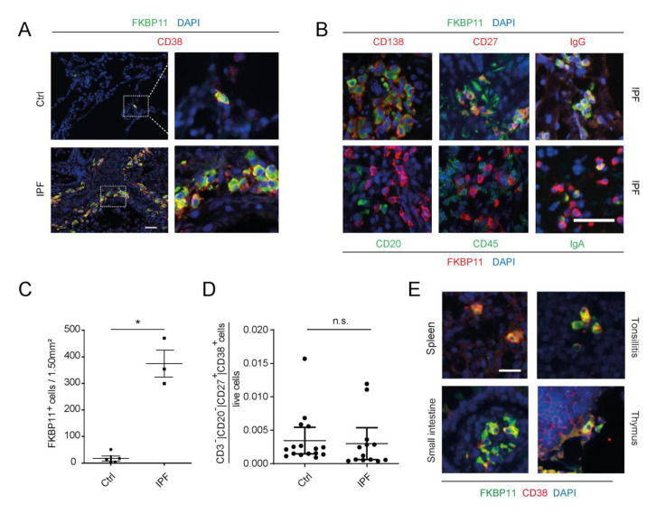
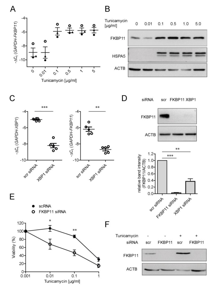
Antibodies are central effectors of the adaptive immune response, widespread used therapeutics, but also potentially disease-causing biomolecules. Antibody folding catalysts in the plasma cell are incompletely defined. Idiopathic pulmonary fibrosis (IPF) is a fatal chronic lung disease with increasingly recognized autoimmune features. We found elevated expression of FK506-binding protein 11 (FKBP11) in IPF lungs where FKBP11 specifically localized to antibody-producing plasma cells. Suggesting a general role in plasma cells, plasma cell-specific FKBP11 expression was equally observed in lymphatic tissues, and in vitro B cell to plasma cell differentiation was accompanied by induction of FKBP11 expression. Recombinant human FKBP11 was able to refold IgG antibody in vitro and inhibited by FK506, strongly supporting a function as antibody peptidyl-prolyl cis-trans isomerase. Induction of ER stress in cell lines demonstrated induction of FKBP11 in the context of the unfolded protein response in an X-box-binding protein 1 (XBP1)-dependent manner. While deficiency of FKBP11 increased susceptibility to ER stress-mediated cell death in an alveolar epithelial cell line, FKBP11 knockdown in an antibody-producing hybridoma cell line neither induced cell death nor decreased expression or secretion of IgG antibody. Similarly, antibody secretion by the same hybridoma cell line was not affected by knockdown of the established antibody peptidyl-prolyl isomerase cyclophilin B. The results are consistent with FKBP11 as a novel XBP1-regulated antibody peptidyl-prolyl cis-trans isomerase and indicate significant redundancy in the ER-resident folding machinery of antibody-producing hybridoma cells.

Keywords: ER stress; FK506-binding protein; antibody folding; immunophilin; interstitial lung disease; lung fibrosis; peptidyl-prolyl isomerase; tacrolimus.


Conflict of interest statement

N.K. (Naftali Kaminski) served as a consultant to Boehringer Ingelheim, Third Rock, Pliant, Samumed, NuMedii, Theravance, LifeMax, Three Lake Partners, Optikira, Astra Zeneca, RohBar, Veracyte, Augmanity, CSL Behring, Galapagos, Gilead and Thyron over the last 3 years, reports Equity in Pliant and Thyron, and a grant from Veracyte, Boehringer Ingelheim, BMS and non-financial support from MiRagen and Astra Zeneca. N.K. (Naftali Kaminski) has IP on novel biomarkers and therapeutics in IPF licensed to Biotech. E.M. received honorarium from Roche, Novartis, Merck, Sanofi, Biogen, and Bioeq and grant support from Novartis, Sanofi, Roche and Merck. O.E. is supported by Bristol Myers Squibb (FP00018992) and serves in advisory capacity to Pieris Pharmaceuticals, Blade Therapeutics, Delta 4 and YAP Therapeutics. L.K. is employee of Secarna Pharmaceuticals, but was still Ph.D. candidate in Staab-Weijnitz Lab when she contributed to the study. The funders had no role in the design of the study; in the collection, analyses, or interpretation of data; in the writing of the manuscript, or in the decision to publish the results. All other authors declare no conflict of interest.

Figures

Figure 1
Figure 1
FKBP11 is upregulated in idiopathic pulmonary fibrosis (IPF). (A) Scatter plot for FKBP gene expression data extracted from microarray data of normal histology control (n = 43) and samples from patients with IPF (n = 99) [50,51]. (B) Western blot analysis of total lung tissue homogenate showed upregulation of FKBP11 in patients with IPF (n = 8) relative to donor samples (n = 5). (C) Densitometric analysis of the Western blot from (B). For Western Blot analysis, data shown are mean ± SEM, and a two-tailed Mann-Whitney test was used for statistical analysis (* p < 0.05). ACTB = β-actin as loading control.
Figure 2
Figure 2
FKBP11 is expressed by CD27+/CD38+/CD138+/CD20/CD45 plasma cells. (A) Immunofluorescent stainings in control (Ctrl, upper panel) and IPF lung tissue sections (lower panel) demonstrate expression of FKBP11 in CD38+ plasma cells. Stainings are representative for n = 5 (Ctrl) and n = 3 (IPF). Scale bar 40 µm. (B) Immunofluorescent stainings in IPF lung tissue sections further demonstrate that FKBP11+ plasma cells are positive for CD138, CD27 (upper panel), but negative for CD20 and CD45 (lower panel) and produce mainly IgG (upper panel, far right), but also IgA (lower panel, far right). Note, that secondary antibody dyes for FKBP11 differ in upper and lower panel. Stainings of IPF lung sections are representative for n = 2 (CD138, CD27, CD20, CD45) and n = 3 (IgG, IgA). Scale bar, 40 µm. (C) Quantification of CD38/FKBP11 immunofluorescent stainings (A), based on lung sections from normal histology controls (n = 5) and IPF patients (n = 3, observer blinded to diagnosis), where FKBP11+/CD38+ cells from ten randomly selected images sized 1.5 mm2 were counted and added up for all 10 images (p = 0.0357, two-tailed Mann-Whitney (D) Numbers of circulating CD20/CD27+/CD38+ plasma cells were not significantly changed (p = 0.2680, two-tailed Mann-Whitney test) between healthy subjects (n = 20) and IPF patients (n = 13). (E) Immunofluorescent stainings of a human tissue array demonstrated FKBP11+/CD38+ cells in other tissues than lung, namely spleen, tonsils, thymus, and small intestine. Scale bar 20 µm. * p < 0.05; n.s., not significant.
Figure 3
Figure 3
In vitro plasma cell differentiation is accompanied by an increase of FKBP11 expression. PBMCs from blood of three healthy volunteers were treated with interleukin-2 (IL2) and R848 to induce differentiation of memory B cells to antibody-producing plasma cells. (A) Increased IgG expression upon treatment was observed using immunofluorescence analysis of cytospins showing cells positive for intracellular IgG. Scale bar 50 µm. (B) IL2/R848 treatment led to increased levels of the B cell activation marker PR domain zinc finger protein 1 (PRDM1). (C,D) Expression of FKBP11 at transcript (C) as well as protein (D) level was increased in the course of B cell to plasma cell differentiation. (D) Upregulation of the ER chaperone HSPA5 confirmed induction of the unfolded protein response (UPR) during B cell differentiation to plasma cells. (E) PBMCs of healthy donors were stimulated for 7 days with IL-2 and R848. Subsequently, the activated and differentiated cells were pre-gated on live and singlet cells and further gated on CD3 and CD19+ B cells (left panel). Plasmablasts (CD19+CD27highCD38high) and non-plasmablast B cells (CD19+CD27lowCD38low) were bulk sorted using FACSAria Fusion (right panel). Flow cytometry panels are displayed from one representative donor of three independent experiments. (F) Expression of FKBP11 and PRDM1 were analyzed by qPCR of the sorted cell fractions. ACTB = β-actin as loading control. For IgG concentrations and qPCR results, data shown are mean ± SEM, and a paired t-test was used for statistical analysis (* p ≤ 0.05; exact p-values: B: p = 0.0474; C: p = 0.0194; F top panel (FKBP11): p = 0.0180; F bottom panel (PRDM1): p = 0.0495).
Figure 4
Figure 4
FKBP11 is induced by ER stress in an XBP1-dependent manner and protects from ER-stress induced cell death. (A,B) Treatment of A549 with the synthetic ER stress inducer tunicamycin led to a dose-dependent increase of FKBP11 expression both on transcript (A) and on protein level (B). Upregulation of the ER chaperone HSPA5 confirmed induction of ER stress. ACTB = β-actin as loading control. (C) Knockdown of XBP1 in A549 cells treated with 0.1 µg/mL tunicamycin was efficient as assessed by qRT-PCR (left, p = 0.0007, paired t-test) and led to a drastic decrease of FKBP11 transcript (right, p = 0.0026, paired t-test). (D) Knockdown of XBP1 equally led to loss of FKBP11 protein (p = 0.0014, paired t-test). This result is shown in comparison to FKBP11 knockdown under similar conditions (p < 0.0001, paired t-test); top panel, representative Western Blot; bottom panel corresponding densitometric analysis). (E) Combining FKBP11 knockdown with different concentrations of tunicamycin ranging from 1 ng/mL to 1 µg/mL and assessing cell viability by trypan blue exclusion showed that FKBP11 knockdown leads to higher susceptibility to ER stress-induced cell death (Exact p-values: 0.01 µg/mL: p = 0.0414; 0.1 µg/mL: p = 0.0068; paired t-test.) (F) Knockdown of FKBP11 in A549 cells was highly efficient in absence and presence of tunicamycin. For (A,E) data shown is based on three and four independent experiments, respectively, and given as mean ± SEM. (B,F) is representative for three independent experiments. For (C,D), data shown is based on five independent experiments and given as mean ± SEM. A paired t-test was used for statistical analysis (* p < 0.05; ** p < 0.01; *** p < 0.001).
Figure 5
Figure 5
Recombinant FKBP11 folds IgG antibody in vitro. (A) Schematic representation of FKBP11 domain structure. For these experiments, the purified FKBP domain of FKBP11 (amino acids G28 - A146) without the N-terminal signal peptide (SigP) and the C-terminal transmembrane region (TM) was used. (B) In vitro antibody refolding kinetics in absence and presence of FKBP11 and recombinant PPIB as positive control. (C) Antibody refolding by FKBP11 was inhibited by tacrolimus (FK506). Data shown is based on three independent experiments and given as mean ± SEM; error bars are missing when they were smaller than the size of the data symbols.
Figure 6
Figure 6
Knockdown of FKBP11 or PPIB does not reduce antibody yield of a hybridoma cell line. (A) Protein levels of FKBP11 and rat IgG in three different rat/mouse hybridoma cell lines (H1–H3) and the mouse myeloma cell line P3X63-Ag8.653 (AG8) used for fusion. H3 was chosen for subsequent experiments. (B) Confocal microscopy demonstrated colocalization of FKBP11 with the ER marker concanavalin A. Scale bar 20 µm. (C) Subcellular fractionation showed a similar enrichment pattern for FKBP11 as for the ER-resident protein calreticulin (CALR), with main localization in the microsomal (ME) and the nuclear extract (NE), but little to no detection in the cytosolic extract (CE) and the chromatin-bound fraction (CB). (D) Representative Western Blot analysis showing levels of FKBP11, the known antibody folding catalyst PPIB, loading control β-actin (ACTB), intracellular IgG antibody heavy (HC) and light chain (LC), and ER chaperone HSPA5, following siRNA-mediated knockdown of FKBP11 or PPIB in hybridoma cell line H3. (E) Mean FKBP11 knockdown efficiency in the rat hybridoma cell line H3 was 78 ± 3% (n = 6; paired t-test; **** p < 0.0001), mean PPIB knockdown efficiency in the same cell line was 60 ± 1% (n = 3; paired t-test; *** p < 0.001). (F) Knockdown of FKBP11 (n = 6) or PPIB (n = 3) did not affect cell viability relative to scr siRNA control. (G) Quantification of immunoblot band intensities (see representative immunoblots for intracellular protein in panel D, for secreted IgG in panel H) following FKBP11 or PPIB knockdown revealed no significant changes except for the siRNA targets FKBP11 and PPIB; data for the latter two is identical to panel E; intrac., intracellular; secr., secreted. (H) Representative Western Blot analysis showing levels of secreted IgG antibody heavy (HC) and light chain (LC), following siRNA-mediated knockdown of FKBP11 or PPIB in hybridoma cell line H3. Quantification is given in panel G (secr. IgG, LC, HC). (I) ELISA-based IgG quantification showed no significant effect on antibody secretion from FKBP11-, PPIB- or scr siRNA-transfected H3 cells (n = 3).

References

    1. Flajnik M.F., Kasahara M. Origin and evolution of the adaptive immune system: Genetic events and selective pressures. Nat. Rev. Genet. 2010;11:47–59. doi: 10.1038/nrg2703. - DOI - PMC - PubMed
    1. Feige M.J., Hendershot L.M., Buchner J. How antibodies fold. Trends Biochem. Sci. 2010;35:189–198. doi: 10.1016/j.tibs.2009.11.005. - DOI - PMC - PubMed
    1. Feige M.J., Buchner J. Principles and engineering of antibody folding and assembly. Biochim. Biophys. Acta. 2014;1844:2024–2031. doi: 10.1016/j.bbapap.2014.06.004. - DOI - PubMed
    1. Moore K.A., Hollien J. The unfolded protein response in secretory cell function. Annu. Rev. Genet. 2012;46:165–183. doi: 10.1146/annurev-genet-110711-155644. - DOI - PubMed
    1. Todd D.J., Lee A.H., Glimcher L.H. The endoplasmic reticulum stress response in immunity and autoimmunity. Nat. Rev. Immunol. 2008;8:663–674. doi: 10.1038/nri2359. - DOI - PubMed

MeSH terms

LinkOut - more resources