. 2022 Apr 15;11(8):1347.
doi: 10.3390/cells11081347.

CFTR Modulation Reduces SARS-CoV-2 Infection in Human Bronchial Epithelial Cells

Affiliations

CFTR Modulation Reduces SARS-CoV-2 Infection in Human Bronchial Epithelial Cells

Virginia Lotti et al. Cells. .

Abstract

People with cystic fibrosis should be considered at increased risk of developing severe symptoms of COVID-19. Strikingly, a broad array of evidence shows reduced spread of SARS-CoV-2 in these subjects, suggesting a potential role for CFTR in the regulation of SARS-CoV-2 infection/replication. Here, we analyzed SARS-CoV-2 replication in wild-type and CFTR-modified human bronchial epithelial cell lines and primary cells to investigate SARS-CoV-2 infection in people with cystic fibrosis. Both immortalized and primary human bronchial epithelial cells expressing wt or F508del-CFTR along with CRISPR/Cas9 CFTR-ablated clones were infected with SARS-CoV-2 and samples were harvested before and from 24 to 72 h post-infection. CFTR function was also inhibited in wt-CFTR cells with the CFTR-specific inhibitor IOWH-032 and partially restored in F508del-CFTR cells with a combination of CFTR modulators (VX-661+VX-445). Viral load was evaluated by real-time RT-PCR in both supernatant and cell extracts, and ACE-2 expression was analyzed by both western blotting and flow cytometry. SARS-CoV-2 replication was reduced in CFTR-modified bronchial cells compared with wild-type cell lines. No major difference in ACE-2 expression was detected before infection between wild-type and CFTR-modified cells, while a higher expression in wild-type compared to CFTR-modified cells was detectable at 72 h post-infection. Furthermore, inhibition of CFTR channel function elicited significant inhibition of viral replication in cells with wt-CFTR, and correction of CFTR function in F508del-CFTR cells increased the release of SARS-CoV-2 viral particles. Our study provides evidence that CFTR expression/function is involved in the regulation of SARS-CoV-2 replication, thus providing novel insights into the role of CFTR in SARS-CoV-2 infection and the development of therapeutic strategies for COVID-19.

Keywords: ACE-2; CFTR; CFTR inhibitor; SARS-CoV-2 virus; cystic fibrosis; human bronchial epithelial cells.


Conflict of interest statement

The authors declare no conflict of interest.

Figures

Figure 1
Figure 1
SARS-CoV-2 replication capability is different in WT and ΔF CFBE41o- cells: (a) Time course measurement of viral load in the supernatant of CFBE41o- cells infected with SARS-CoV-2 by commercial qualitative real-time RT-PCR (Seegene) able to detect several viral gene targets. The data are presented as the mean ± SD from independent experiments (n = 4; *** p < 0.001). (b) Measurement of ORF-1ab mRNA expression normalized to GAPDH (used as a housekeeping control) in cells infected with RNA extracted from SARS-CoV-2-infected CFBE41o- cells. The data are presented as the mean ± SD from independent experiments (n = 3; * p < 0.05, ** p < 0.01). (c) Time course measurement of viral load in the supernatant of WT and ΔF CFBE41o- cells infected with the BetaCoV/Australia/VIC01/2020 SARS-CoV-2 strain. The data were obtained from semiquantitative PCR (Seegene) of cellular supernatant and are expressed as the copies/µL ratio. (d) Total S1 protein quantification by Western blot analysis in CFBE41o- cells. Samples analyzed were harvested from 0 to 72 hpi with SARS-CoV-2. β-Actin was used as a loading control. The blot is representative of n = 3.
Figure 2
Figure 2
ACE-2 expression levels in CFBE41o- cells determined by different techniques: (a) Flow cytometry of ACE-2 cell membrane expression in uninfected ΔF and WTCFBE41o- cells. The data are representative of n = 3. (b) MFI of ACE-2 in uninfected ΔF and WT CFBE41o- cells. The data are presented as the mean ± SD from independent experiments (n = 3). (c) Total ACE-2 protein quantification by Western blot analysis in CFBE41o- cells. The samples analyzed were harvested from 0 to 72 hpi with SARS-CoV-2. β-Actin was used as a loading control. The data are from a Western blot representative of n = 3. (d) Measurement of ACE-2 mRNA expression normalized to GAPDH (used as a housekeeping control) in RNA extracted from SARS-CoV-2-infected CFBE41o- cells. The data are presented as the mean ± SD from independent experiments (n = 3).
Figure 3
Figure 3
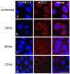
Immunofluorescence confocal microscopy showing the expression of ACE-2 (red) in uninfected (a) and infected CFBE41o- WT cells obtained at 24 (b), 48 (c), 72 (d) hpi. Cell nuclei were counterstained with TO-PRO 3 (blue). Scale bar: 20 µm.
Figure 4
Figure 4
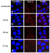
Immunofluorescence confocal microscopy showing the expression of ACE-2 (red) in uninfected (a) and infected CFBE41o- ΔF cells obtained at 24 (b), 48 (c), 72 (d) hpi. Cell nuclei were counterstained with TO-PRO 3 (blue). Scale bar: 20 µm.
Figure 5
Figure 5
SARS-CoV-2 replication and ACE-2 expression in WT and KO 16HBE14o- cells: (a) Time course measurement of viral load in the supernatant of 16HBE14o- cells infected with SARS-CoV-2 analyzed by qualitative real-time RT-PCR (Seegene) able to detect several viral gene targets. The data are presented as the mean ± SD from independent experiments (n = 4; ** p < 0.01). (b) Flow cytometry analysis of ACE-2 cell membrane expression in uninfected WT and KO 16HBE14o- cells. The data are representative of n = 3 measurements. (c) MFI of ACE-2 in uninfected WT and KO 16HBE14o- cells. The bars represent the mean ± SD from independent experiments (n = 3; * p < 0.05). (d) Total ACE-2 protein quantification by Western blot analysis in 16HBE14o- cells. The samples analyzed were harvested from 0 to 72 hpi with SARS-CoV-2. β-Actin was used as a loading control. The data are representative of n = 3.
Figure 6
Figure 6
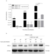
Effect of CFTR inhibition in CFBE41o- WT cells via IOWH-032 treatment compared to WT and ΔF CFBE41o- cells: (a) Time course measurement of viral load in the supernatant of CFBE41o- cells infected with SARS-CoV-2 analyzed by qualitative real-time RT-PCR (Seegene). The data are presented as the mean ± SD from independent experiments (n = 3; **** p < 0.0001). (b) Total ACE-2 protein quantification by Western blot analysis in CFBE41o- cells. Samples were harvested from CFBE41o- WT cells exposed or not to IOWH-032 as well as CFBE41o- ΔF cells at different time points of SARS-CoV-2 infection. β-Actin was used as a loading control. The data are representative of n = 3 experiments.
Figure 7
Figure 7
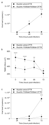
SARS-CoV-2 replication in wt/wt-CFTR and F508del/F508del-CFTR MucilAir™: (a) Apical viral production was assessed in washes of the apical side at 24, 48, 72 hpi of both wt/wt-CFTR and F508del/F508del-CFTR MucilAir™ by commercial qualitative real-time RT-PCR (Seegene). The data are presented as the mean ± SD from independent experiments (n = 3; ** p < 0.01; **** p < 0.0001). (b) Trans-epithelial resistance measurements in wt/wt-CFTR and F508del/F508del-CFTR MucilAir™ (TEER in Ω·cm2) between the apical and basal sides at each time point. The data are presented as the mean ± SD from independent experiments (n = 3). (c) Basolateral SARS-CoV-2 quantification in wt/wt-CFTR and F508del/F508del-CFTR MucilAir™. Basolateral SARS-CoV-2 viral quantification was assessed in the basolateral medium at 24, 48, 72 hpi of both wt/wt-CFTR and F508del/F508del-CFTR MucilAir™ by commercial qualitative real-time RT-PCR (Seegene) able to detect several viral gene targets. The data are presented as the mean ± SD from independent experiments (n = 3; **** p < 0.0001).
Figure 8
Figure 8
SARS-CoV-2 replication cycle in MucilAir™ is driven by CFTR modulation: (a) Apical viral production was assessed in washes of the apical side at 24, 48, 72 hpi of both wt/wt-CFTR and F508del/F508del-CFTR MucilAir™ with and without CFTR-modulator/inhibitor treatment by commercial qualitative real-time RT-PCR (Seegene). The data are presented as the mean ± SD from independent experiments (n = 3; * p < 0.05; ** p < 0.01; *** p < 0.001; **** p < 0.0001). (b) Trans-epithelial resistance (TEER in Ω·cm2) between the apical and basal sides was measured at each time point in both wt/wt-CFTR and F508del/F508del-CFTR MucilAir™ with and without VX-661+VX-445, for F508del/F508del-CFTR, or IOWH-032, for wt/wt-CFTR, chronic treatments. The data are presented as the mean ± SD from independent experiments (n = 3).

References

    1. Zhu N., Zhang D., Wang W., Li X., Yang B., Song J., Zhao X., Huang B., Shi W., Lu R., et al. A Novel Coronavirus from Patients with Pneumonia in China, 2019. N. Engl. J. Med. 2020;382:727–733. doi: 10.1056/NEJMoa2001017. - DOI - PMC - PubMed
    1. Huang C., Wang Y., Li X., Ren L., Zhao J., Hu Y., Zhang L., Fan G., Xu J., Gu X., et al. Clinical features of patients infected with 2019 novel coronavirus in Wuhan, China. Lancet. 2020;395:497–506. doi: 10.1016/S0140-6736(20)30183-5. - DOI - PMC - PubMed
    1. Hui D.S., Ahzar E.I., Madani T.A., Ntoumi F., Kock R., Dar O., Ippolito G., McHugh T.D., Memish Z.A., Drosten C., et al. The continuing 2019-nCoV epidemic threat of novel coronaviruses to global health—The latest 2019 novel coronavirus outbreak in Wuhan, China. Int. J. Infect. Dis. 2020;91:264–266. doi: 10.1016/j.ijid.2020.01.009. - DOI - PMC - PubMed
    1. W.H.O WHO Director-General’s Opening Remarks at the Media Briefing on COVID-19. 2020. [(accessed on 11 March 2020)]. Available online: https://www.who.int/dg/speeches/detail/who-director-general-s-opening-re....
    1. Hoffmann M., Kleine-Weber H., Schroeder S., Kruger N., Herrler T., Erichsen S., Schiergens T.S., Herrler G., Wu N.H., Nitsche A., et al. SARS-CoV-2 Cell Entry Depends on ACE2 and TMPRSS2 and Is Blocked by a Clinically Proven Protease Inhibitor. Cell. 2020;181:271–280.e8. doi: 10.1016/j.cell.2020.02.052. - DOI - PMC - PubMed

MeSH terms

Substances