. 2022 Apr 15;11(8):1357.
doi: 10.3390/cells11081357.

Rank-Rankl-Opg Axis in Multiple Sclerosis: The Contribution of Placenta

Affiliations

Rank-Rankl-Opg Axis in Multiple Sclerosis: The Contribution of Placenta

Sofia Passaponti et al. Cells. .

Abstract

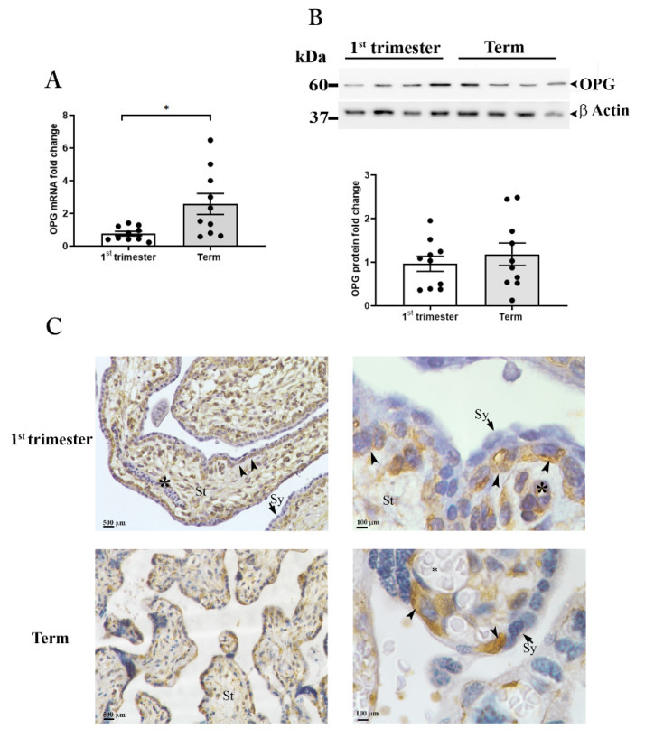
Women with multiple sclerosis (MS) can safely become pregnant and give birth, with no side effects or impediments. Pregnancy is generally accepted as a period of well-being in which relapses have a softer evolution, particularly in the third trimester. Herein, we hypothesized that the placenta, via its "secretome", could contribute to the recognized beneficial effects of pregnancy on MS activity. We focused on a well-known receptor/ligand/decoy receptor system, such as the one composed by the receptor activator of nuclear factor-kB (RANK), its ligand (RANKL), and the decoy receptor osteoprotegerin (OPG), which have never been investigated in an integrated way in MS, pregnancy, and placenta. We reported that pregnancy at the term of gestation influences the balance between circulating RANKL and its endogenous inhibitor OPG in MS women. We demonstrated that the placenta at term is an invaluable source of homodimeric OPG. By functional studies on astrocytes, we showed that placental OPG suppresses the mRNA expression of the CCL20, a chemokine responsible for Th17 cell recruitment. We propose placental OPG as a crucial molecule for the recognized beneficial effect of late pregnancy on MS and its potential utility for the development of new and more effective therapeutic approaches.

Keywords: autoimmune diseases; osteoprotegerin; placenta; pregnancy.


Conflict of interest statement

The authors declare no conflict of interest. The funders had no role in the design of the study; in the collection, analyses, or interpretation of data; in the writing of the manuscript, or in the decision to publish the results.

Figures

Figure 1
Figure 1
OPG and sRANKL in serum samples OPG and sRANKL in serum of MS women (A,B): ELISA assay for OPG (A) and sRANKL (B) in serum from non-pregnant women of reproductive age (NP; n = 19), at term of gestation (Term; n = 10) and in the post-partum period (PP; n = 10). OPG and sRANKL in serum of healthy women (C,D): ELISA assay for OPG (C) and sRANKL (D) in serum from healthy non-pregnant women of reproductive age (NP; n = 10), pregnant women, at first-trimester (1st Trimester; n = 10) and term of gestation (Term; n = 20). Serum sRANKL/OPG ratio in MS and healthy women (E): sRANKL/OPG ratio for MS patients, (non-pregnant: NP, n = 19; at term of gestation: Term, n = 10) and healthy women (non-pregnant: NP, n = 10; at term of gestation: Term, n = 20). Red circle in MS at term identifies one outlier. OPG quantification in paired maternal and cord serum (F): ELISA assay for OPG levels in paired maternal and cord serum (n = 10 donors). Each symbol represents a unique donor and mean ± SEM is shown for data (AD) and (F). Data are presented as box-whisker plots in (E). Significance was determined with the Kruskal–Wallis test for multiple comparisons for data (AE) and paired t-test for data in (F). * p < 0.05; ** p < 0.01; *** p < 0.001.
Figure 2
Figure 2
OPG expression in placental tissues. OPG mRNA expression (A) in placenta tissues at first-trimester (1st trimester; n = 10) and term (n = 10) pregnancy. (B) Representative Western blot and relative OPG protein expression in first-trimester (1st Trimester; n = 10) and term (Term; n = 10) placenta tissues. β-actin was used as loading control. (C) Representative OPG immunolocalization in first-trimester (upper panels) and term placenta tissues (lower panels) (n = 3 for each gestational period); villous stroma (St), perivascular cells of fetal capillaries (asterisk), cytotrophoblasts (arrowhead), syncytiotrophoblast (Sy, arrows). Data are presented as individual values, mean ± SEM are shown. Significance was determined with the unpaired t-test. * p < 0.05.
Figure 3
Figure 3
OPG production by placenta explants. ELISA assay for OPG in conditioned media (A) from first-trimester (maintained at 3% of oxygen for 48 h; n = 4 placentae at 9–11 weeks) and term (maintained at 8% of oxygen; n = 6 placentae at 38–39 weeks) placenta explants. Representative Western blot (B) under non-reducing conditions for OPG in conditioned media from first-trimester and term placenta explants. ELISA assay for OPG in conditioned media (C) from term placenta explants (maintained at 8% of oxygen; n = 3 placentae at 38–39 weeks) exposed for 48 h to estradiol (E2, 1 × 10⁻⁸ M), estriol (E3 7 × 10⁻⁸ M) and progesterone (Pg, 1 × 10⁻⁶ M) or medium alone (control, Ct). Data expressed as mean ± SEM. Significance was determined with the Unpaired t-test for OPG production in basal condition and one-way ANOVA for hormones exposure experiments. * p < 0.05; ** p < 0.01; *** p < 0.001.
Figure 4
Figure 4
RANK-RANKL-OPG in human astrocytes. Representative immunofluorescence staining for RANK in astrocytes (A). RANK protein expression in astrocytes after the treatment with rh RANKL (original magnification 63X) (B). mRNA quantification for cytokines and chemokines in rh RANKL treated astrocytes (C). mRNA quantification for CCL20 in astrocytes treated with rh RANKL plus Conditioned Media (CM) from term placenta explants or CM OPG-depleted (D). mRNA quantification for CCL20 in astrocytes treated with rh RANKL or RANKL plus rh OPG (25, 50 and 100 ng/mL) (E). Data expressed as mean ± SEM of four separate experiments performed in quadruplicate. Significance was determined with Mann–Whitney test for data in (C) and one-way ANOVA for data in (D,E). * p < 0.05; ** p < 0.01.
Figure 5
Figure 5
Schematic representation of the effect of pregnancy on sRANKL to OPG ratio in multiple sclerosis patients. MS patients have a dysregulated sRANKL to OPG ratio and increased expression of CCL20 within the CNS. During the third trimester of pregnancy, the placenta releases OPG in the maternal circulation. Placental OPG reverts the sRANKL/OPG balance, exerts inhibition on RANK-RANKL interaction within the maternal CNS, and leads to a reduction of CCL20 by astrocytes.

References

    1. Confavreux C., Hutchinson M., Hours M.M., Cortinovis-Tourniaire P., Moreau T. Rate of pregnancy-related relapse in multiple sclerosis. Pregnancy in Multiple Sclerosis Group. N. Engl. J. Med. 1998;339:285–291. doi: 10.1056/NEJM199807303390501. - DOI - PubMed
    1. Dendrou C.A., Fugger L., Friese M.A. Immunopathology of multiple sclerosis. Nat. Rev. Immunol. 2015;15:545–558. doi: 10.1038/nri3871. - DOI - PubMed
    1. Voskuhl R.R., Gold S.M. Sex-related factors in multiple sclerosis susceptibility and progression. Nat. Rev. Neurol. 2012;8:255–263. doi: 10.1038/nrneurol.2012.43. - DOI - PMC - PubMed
    1. Roux T., Courtillot C., Debs R., Touraine P., Lubetzki C., Papeix C. Fecundity in women with multiple sclerosis: An observational mono-centric study. J. Neurol. 2015;262:957–960. doi: 10.1007/s00415-015-7663-1. - DOI - PubMed
    1. Arafa A., Wang X., Dong J.Y., Eshak E.S. Does multiple sclerosis increase the risk of preeclampsia? A systematic review and meta-analysis. Hypertens Pregnancy. 2021;40:180–185. doi: 10.1080/10641955.2021.1921792. - DOI - PubMed

Publication types