. 2022 Apr 16;11(8):1361.
doi: 10.3390/cells11081361.

Verteporfin Inhibits the Progression of Spontaneous Osteosarcoma Caused by Trp53 and Rb1 Deficiency in Ctsk-Expressing Cells via Impeding Hippo Pathway

Affiliations

Verteporfin Inhibits the Progression of Spontaneous Osteosarcoma Caused by Trp53 and Rb1 Deficiency in Ctsk-Expressing Cells via Impeding Hippo Pathway

Yang Li et al. Cells. .

Abstract

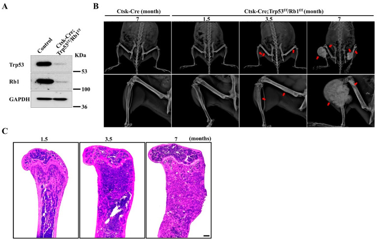
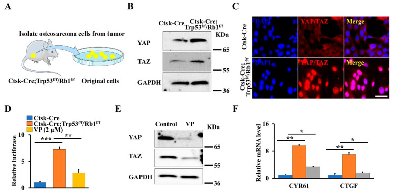
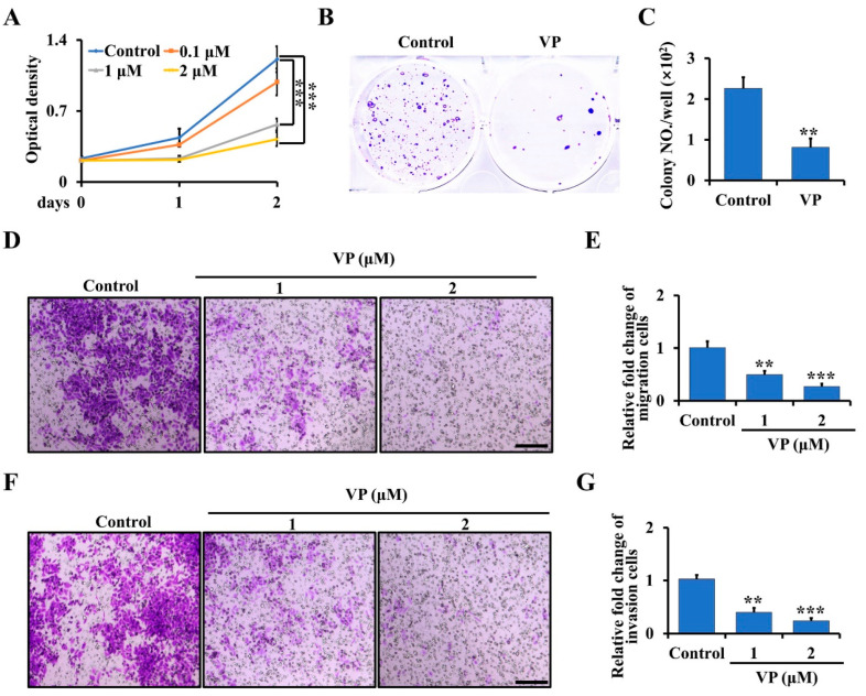
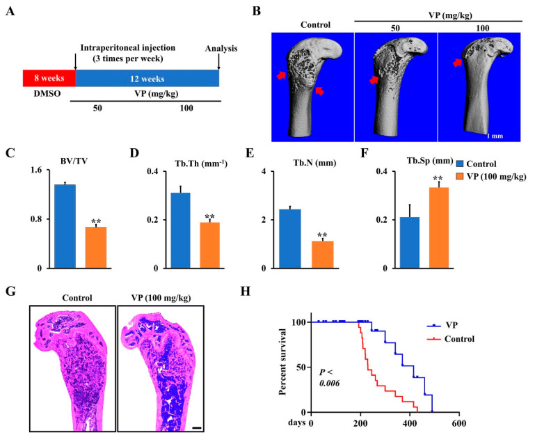
Osteosarcoma is the most common primary malignancy of bone in children and adolescents. Others and our previous studies have shown that Yes-associated protein (YAP)/transcriptional coactivator with PDZ-binding motif (TAZ) as core components of the Hippo pathway are crucial regulators of osteosarcoma formation and progression. Recent studies demonstrated that verteporfin (VP) is an inhibitor of YAP/TAZ signaling in xenograft osteosarcoma. However, whether VP can inhibit primary osteosarcoma in mice is unknown. Mutations of Trp53 and Rb1 occur in approximately 50~70% of human osteosarcoma. In this study, we successfully generated the Ctsk-Cre;Trp53f/f/Rb1f/f mice in which Trp53/Rb1 was ablated in Ctsk-expressing cells and found that Ctsk-Cre;Trp53f/f/Rb1f/f mice spontaneously developed osteosarcoma with increased expansive osteoid lesions in the cortical bone with aging. Loss of Trp53/Rb1 in Ctsk-expressing cells significantly promoted the expression and nuclear translocation of YAP/TAZ. Micro-CT results showed that inhibition of YAP/TAZ by VP delays osteosarcoma progression and protected against bone erosion in Ctsk-Cre;Trp53f/f/Rb1f/f mice. Importantly, the Kaplan-Meier survival curves displayed a significantly longer survival rate after VP treatment in Ctsk-Cre;Trp53f/f/Rb1f/f mice compared to non-injected groups. In vitro studies further showed that VP inhibited the proliferation, migration and invasion in Trp53/Rb1-mutant Ctsk-expressing cells. Moreover, the results from promoter luciferase activity analysis showed that the transcriptional activity of YAP/TAZ was significantly increased in osteosarcoma tissue from Ctsk-Cre;Trp53f/f/Rb1f/f mice, which was attenuated by VP treatment. Overall, these findings suggest that targeting Hippo pathway by VP may be a potential therapeutic strategy for osteosarcoma.

Keywords: Rb1; Trp53; YAP/TAZ; osteosarcoma; verteporfin.


Conflict of interest statement

The authors in this study declare no competing interest.

Figures

Figure 1
Figure 1
Loss of Trp53 and Rb1 in Ctsk-expressing cells causes osteosarcoma formation. (A) Western blot analysis of Trp53 and Rb1 expressions in Ctsk-expressing cells and normal cortical bone cells (control) as indicated. (B) Representative X-ray images of Ctsk-Cre;Trp53f/f/Rb1f/f mice and controls as indicated timepoint. The red arrows indicate the tumor in the bone. N = 5 mice per group. (C) Representative H&E staining images of femurs from the Ctsk-Cre;Trp53f/f/Rb1f/f mice as indicated timepoints. Scale bar, 1 mm. N = 5 mice per group.
Figure 2
Figure 2
Loss of Trp53 and Rb1 in Ctsk-expression activates YAP/TAZ signaling. (A) Schematic presentation of primary osteosarcoma cells from Ctsk-Cre;Trp53f/f/Rb1f/f mice. (B) Western blot analysis of YAP and TAZ expression in primary osteosarcoma cells and normal cortical bone cells. (C) Representative images of immunofluorescent staining of YAP/TAZ in the primary osteosarcoma cells and normal cortical bone cells as indicated. Scale bars, 10 μm. (D) The normal cortical bone cells or primary osteosarcoma cells with/without 2 μmol VP were co-transfected with 8xGTIIC and pRL-TK plasmids (internal control) as indicated, respectively. After transfection of 48 h, the luciferase activities were identified by the Dual-Luciferase Assay Kit. (E) The primary osteosarcoma cells were treated by 2 μmol VP or DMSO (control) for 48 h, the protein levels of YAP and TAZ were identified as indicated. (F) qRT-PCR analysis of YAP/TAZ target genes CYR61 and CTGF in the normal cortical bone cells or primary osteosarcoma cells cultured with/without 2 μmol VP for 48 h as indicated. Error bars were the means ± SEM from three independent experiments. * p < 0.05. ** p < 0.01, *** p < 0.001.
Figure 3
Figure 3
VP inhibits proliferation, migration, and invasion in Trp53/Rb1-mutant Ctsk-expressing osteosarcoma cells. (A) The primary osteosarcoma cells from Ctsk-Cre;Trp53f/f/Rb1f/f mice were collected and seeded in a 96-well plate. After being cultured for the indicated time, the cell proliferation was determined by WST-1 Cell Proliferation Assay Kit. (B,C) Soft agar assay (B). The primary osteosarcoma cells from Ctsk-Cre;Trp53f/f/Rb1f/f mice were incubated with 0 or 2 μM VP for 3 weeks, and then the colony numbers were counted (C). (D,E) The primary osteosarcoma cells were isolated from Ctsk-Cre;Trp53f/f/Rb1f/f mice, and treated with indicated doses of VP in transwell plates. After incubation for 48 h, the migrated cells were stained by crystal violet and viewed under microscope (D). Scale bars, 100 μm. The corresponding quantification was identified (E). (F,G) Cell invasion and quantification as indicated. Scale bars, 100 μm. Error bars were the means ± SEM from three independent experiments. ** p < 0.01, *** p < 0.001.
Figure 4
Figure 4
Inhibition of Hippo pathway by VP inhibits osteosarcoma progression in Ctsk-Cre;Trp53f/f/Rb1f/f mice. (A) Schematic diagram of the time courses of VP injection and mice harvested for data analysis. (B) Representative X-ray images of femurs showing that treatment of Ctsk-Cre;Trp53f/f/Rb1f/f mice with VP 3 times per week for 12 weeks starting from 2 months. Scale bars, 1 mm. The red arrows indicate the destruction in the cortical bone. (CF) Histomorphometric analysis of bone parameters in the femurs of Ctsk-Cre;Trp53f/f/Rb1f/f mice after treatment with 0 (control) or 100 mg/kg VP as indicated. N = 5 mice/group. (G) Representative H&E-stained images of femurs from Ctsk-Cre;Trp53f/f/Rb1f/f mice treated with 0 (control) or 100 mg/kg VP for 12 weeks. Scale bar, 1 mm. (H) Kaplan–Meier survival analysis indicating overall survival of treated with 0 (control) or 100 mg/kg VP for 12 weeks. Error bars were the means ± SEM from three independent experiments. ** p < 0.01.

References

    1. Li Y., Liu M., Yang S., Fuller A.M., Karin Eisinger-Mathason T.S., Yang S. RGS12 is a novel tumor suppressor in osteosarcoma that inhibits YAP-TEAD1-Ezrin signaling. Oncogene. 2021;40:2553–2566. doi: 10.1038/s41388-020-01599-z. - DOI - PMC - PubMed
    1. Pan D.J. The Hippo Signaling Pathway in Development and Cancer. Dev. Cell. 2010;19:491–505. doi: 10.1016/j.devcel.2010.09.011. - DOI - PMC - PubMed
    1. Kegelman C.D., Mason D.E., Dawahare J.H., Horan D.J., Vigil G.D., Howard S.S., Robling A.G., Bellido T.M., Boerckel J.D. Skeletal cell YAP and TAZ combinatorially promote bone development. Faseb J. 2018;32:2706–2721. doi: 10.1096/fj.201700872R. - DOI - PMC - PubMed
    1. Harvey K.F., Zhang X., Thomas D.M. The Hippo pathway and human cancer. Nat. Rev. Cancer. 2013;13:246–257. doi: 10.1038/nrc3458. - DOI - PubMed
    1. Zheng Y., Pan D. The Hippo Signaling Pathway in Development and Disease. Dev. Cell. 2019;50:264–282. doi: 10.1016/j.devcel.2019.06.003. - DOI - PMC - PubMed

Publication types