

Role of Lung Ultrasound in the Management of Patients with Suspected SARS-CoV-2 Infection in the Emergency Department
- PMID: 35456160
- PMCID: PMC9025104
- DOI: 10.3390/jcm11082067
Role of Lung Ultrasound in the Management of Patients with Suspected SARS-CoV-2 Infection in the Emergency Department
Abstract
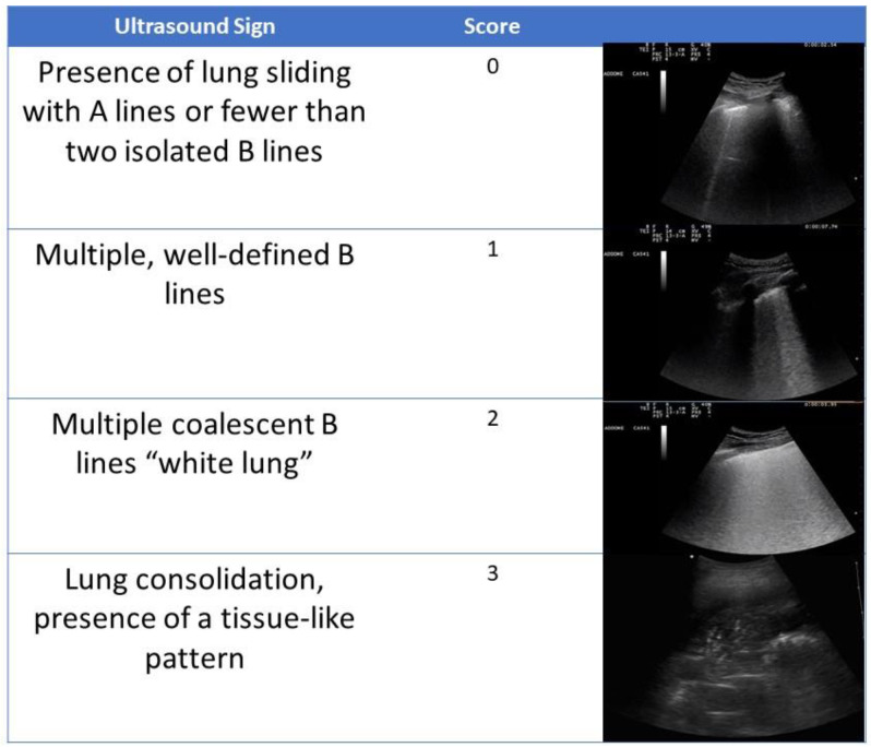
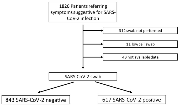
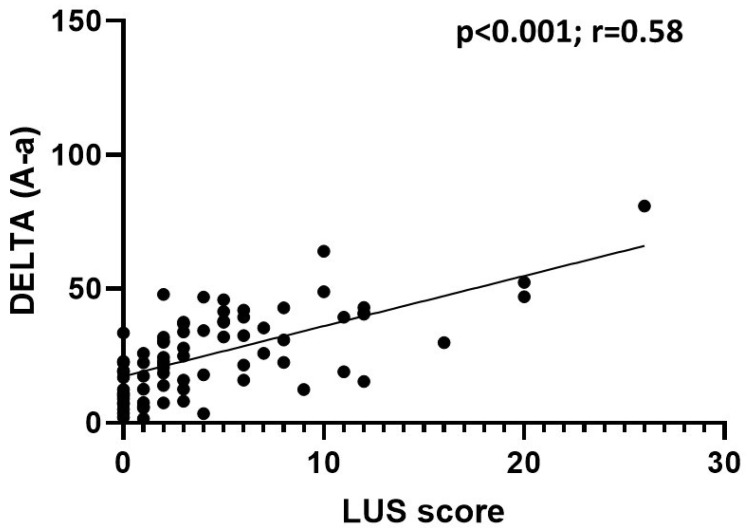
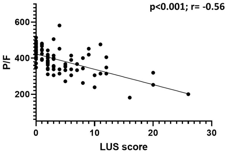
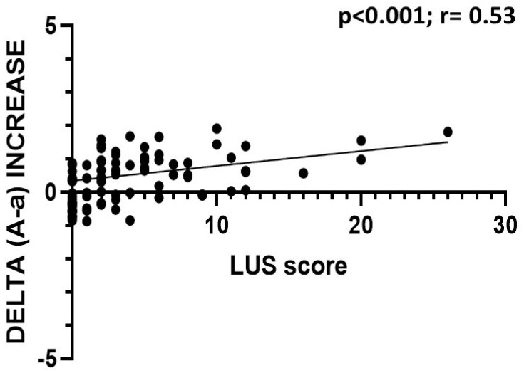
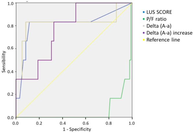
Background: The lung ultrasound (LUS) score has been proposed as an optimal scheme for the ultrasound study of patients with suspected/confirmed COVID-19 pneumonia. The aims of our study were to evaluate the use of lung ultrasound as a diagnostic tool for diagnosing SARS-CoV-2 pneumonia, to examine the validity of the LUS score for the diagnosis of COVID-19 pneumonia, and to correlate this score with hospitalization rate and 30-day mortality. Materials and Methods: A retrospective analysis was performed on 1460 patients who were referred to the General Emergency Department of the S. Orsola-Malpighi Hospital from April 2020 to May 2020 for symptoms suspected to indicate SARS-CoV-2 infection. The ultrasound examination was based on a common execution scheme called the LUS score, as previously described. Results and Conclusions: The LUS score was found to correlate with the degree of clinical severity and respiratory failure (paO2/FiO2 ratio and the alveolar−arterial gradient increase than expected for age). It was shown that COVID-19 patients with an LUS score of >7 require the use of oxygen support, and a value of >10 is associated with an increased risk of oro-tracheal intubation. The LUS score was found to present higher values in hospitalized patients, increasing according to the degree of care intensity. Patients who died from COVID-19 were characterized by a mean LUS score of 11 at presentation to the emergency department. An LUS score of >7.5 was found to indicate a sensitivity of 83% and a specificity of 89% for 30-day mortality in COVID-19 patients. The use of LUS seems to be an optimal first level method for pneumonia detection and risk stratification in patients with suspected SARS-CoV-2 infection.
Keywords: COVID-19; SARS-CoV-2; infection; lung; pneumonia; ultrasound.
Conflict of interest statement
The authors declare they have no conflict of interest.
Figures
Similar articles
-
Lung ultrasound in the emergency department - a valuable tool in the management of patients presenting with respiratory symptoms during the SARS-CoV-2 pandemic.BMC Emerg Med. 2020 Dec 7;20(1):96. doi: 10.1186/s12873-020-00389-w. BMC Emerg Med. 2020. PMID: 33287732 Free PMC article.
-
Comparative study of lung ultrasound and chest computed tomography scan in the assessment of severity of confirmed COVID-19 pneumonia.Intensive Care Med. 2020 Sep;46(9):1707-1713. doi: 10.1007/s00134-020-06186-0. Epub 2020 Jul 29. Intensive Care Med. 2020. PMID: 32728966 Free PMC article.
-
Lung ultrasound as diagnostic tool for SARS-CoV-2 infection.Intern Emerg Med. 2021 Mar;16(2):471-476. doi: 10.1007/s11739-020-02512-y. Epub 2020 Oct 3. Intern Emerg Med. 2021. PMID: 33011929 Free PMC article.
-
Lung Ultrasound Score in COVID-19 Patients Correlates with PO2/FiO2, Intubation Rates, and Mortality.West J Emerg Med. 2024 Jan;25(1):28-39. doi: 10.5811/westjem.59975. West J Emerg Med. 2024. PMID: 38205982 Free PMC article.
-
Lung ultrasound score severity cut-off points in COVID-19 pneumonia. A systematic review and validating cohort.Med Clin (Barc). 2023 Jun 23;160(12):531-539. doi: 10.1016/j.medcli.2023.01.024. Epub 2023 Mar 10. Med Clin (Barc). 2023. PMID: 36990898 Free PMC article.
Cited by
-
Role of Lung Ultrasound in the Detection of Lung Sequelae in Post-COVID-19 Patients: A Systematic Review and Meta-Analysis.J Clin Med. 2024 Sep 21;13(18):5607. doi: 10.3390/jcm13185607. J Clin Med. 2024. PMID: 39337096 Free PMC article. Review.
-
Ultrasound diagnosis of a pseudoaneurysm of the internal right mammary artery.J Ultrasound. 2024 Jun;27(2):393-396. doi: 10.1007/s40477-024-00889-6. Epub 2024 Apr 17. J Ultrasound. 2024. PMID: 38632171 Free PMC article.
-
New perspectives on the use of artificial intelligence in the ultrasound evaluation of lung diseases.J Ultrasound. 2024 Jun;27(2):429-431. doi: 10.1007/s40477-023-00866-5. Epub 2024 Feb 5. J Ultrasound. 2024. PMID: 38315408 Free PMC article. No abstract available.
-
Comparison of positive pressure extubation with traditional extubation in critically ill patients - a randomised control study.Anaesthesiol Intensive Ther. 2023;55(1):38-45. doi: 10.5114/ait.2023.125584. Anaesthesiol Intensive Ther. 2023. PMID: 37306270 Free PMC article. Clinical Trial.
-
The correlation between lung ultrasound scores and outcomes of high-flow nasal cannula therapy in infants with severe pneumonia.BMC Pediatr. 2024 Jan 16;24(1):51. doi: 10.1186/s12887-024-04522-7. BMC Pediatr. 2024. PMID: 38229006 Free PMC article.
References
-
- Soldati G., Smargiassi A., Inchingolo R., Buonsenso D., Perrone T., Briganti D.F., Perlini S., Torri E., Mariani A., Mossolani E.E., et al. On Lung Ultrasound Patterns Specificity in the Management of COVID-19 Patients. J. Ultrasound Med. 2020;39:2283–2284. doi: 10.1002/jum.15326. - DOI - PMC - PubMed
-
- Guarracino F., Vetrugno L., Forfori F., Corradi F., Orso D., Bertini P., Ortalda A., Federici N., Copetti R., Bove T. Lung, Heart, Vascular, and Diaphragm Ultrasound Examination of COVID-19 Patients: A Comprehensive Approach. J. Cardiothorac. Vasc. Anesth. 2020;35:1866–1874. doi: 10.1053/j.jvca.2020.06.013. - DOI - PMC - PubMed
LinkOut - more resources
Full Text Sources
Miscellaneous
