Review
. 2022 Apr 8;11(8):2094.
doi: 10.3390/jcm11082094.

Genetic Insights into Primary Restrictive Cardiomyopathy

Affiliations
Review

Genetic Insights into Primary Restrictive Cardiomyopathy

Andreas Brodehl et al. J Clin Med. .

Abstract

Restrictive cardiomyopathy is a rare cardiac disease causing severe diastolic dysfunction, ventricular stiffness and dilated atria. In consequence, it induces heart failure often with preserved ejection fraction and is associated with a high mortality. Since it is a poor clinical prognosis, patients with restrictive cardiomyopathy frequently require heart transplantation. Genetic as well as non-genetic factors contribute to restrictive cardiomyopathy and a significant portion of cases are of unknown etiology. However, the genetic forms of restrictive cardiomyopathy and the involved molecular pathomechanisms are only partially understood. In this review, we summarize the current knowledge about primary genetic restrictive cardiomyopathy and describe its genetic landscape, which might be of interest for geneticists as well as for cardiologists.

Keywords: cardiomyopathy; cardiovascular genetics; desmin; filamin-C; restrictive cardiomyopathy; troponin.


Conflict of interest statement

The authors declare no conflict of interest. The funders had no role in the design of the study; in the collection, analyses, or interpretation of data; in the writing of the manuscript, or in the decision to publish the results.

Figures

Figure 4
Figure 4
Schematic molecular structure of the thin filaments in the Ca2+ free state [68] (https://www.rcsb.org/structure/6KN7 (accessed on 13 March 2022)). Actin is shown in light green, tropomyosin is shown in red, cardiac troponin T is shown in blue, troponin C is shown in violet and troponin I is shown in orange. The localizations of the RCM-associated TNNI3 missense mutations are shown in cyan. The majority of RCM-associated TNNI3 missense mutations are localized in the C-terminal part of troponin-I.
Figure 6
Figure 6
Structural overview of the anti parallel α-actinin-2 dimer (https://www.rcsb.org/structure/4D1E) (accessed on 13 March 2022) [149]. The N-terminal. Actin-binding domains are shown in red. Four spectrin-like repeats build the central cylindrical rod domain (green). A C-terminal calmodulin-like domain is built by two EF hand motifs (purple and blue). The position of the RCM-associated mutation ACTN2-p.N157Y within the actin-binding domain is shown in cyan.
Figure 1
Figure 1
(A) Apical four chamber view during systole of an echocardiogram (B) and four chamber view of cardiac magnetic resonance image of a 50-year-old patient carrying a pathogenic FLNC mutation. Note the enlarged atria, normal ventricular sizes and wall thicknesses. RA = right atrium; RV = right ventricle; LA = left atrium; LV = left ventricle.
Figure 2
Figure 2
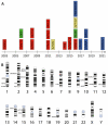
Overview of RCM genes. (A) Genes associated with restrictive cardiomyopathy (RCM) according to the year of discovery. Different subcellular localizations are color-coded (red = sarcomere; green = cytoskeleton; yellow = Z-disc and blue = others). (B) Chromosomal location of RCM-associated genes. Schematic idiograms were licensed from shutterstock.de.
Figure 3
Figure 3
Venn diagram showing the genetic overlap of restrictive cardiomyopathy (RCM) with other cardiomyopathies. ACM = arrhythmogenic cardiomyopathy; DCM = dilated cardiomyopathy; HCM = hypertrophic cardiomyopathy; LVNC = left ventricular non-compaction cardiomyopathy; and MFM = myofibrillar myopathy. Gene names according to the HUGO Gene Nomenclature Committee, HGNC (https://www.genenames.org/ (accessed on 13 March 2022)). Sub-images of the DCM or HCM heart were licensed from shutterstock.de.
Figure 5
Figure 5
Schematic overview of RCM associated DES mutations. (A) Schematic overview about the DES gene consisting of nine exons (NM_001927.4). Three splice site mutations have been identified in RCM patients at the donor splice site of exon 3. (B) Schematic domain organization of desmin and the localization of the known RCM-associated DES missense mutations.
Figure 7
Figure 7
Schematic overview about the domain organization of filamin-C and the localization of the known RCM-associated FLNC missense mutations.
Figure 8
Figure 8
Molecular structure of the αB-crystallin domain determined by nuclear magnetic resonance (NMR) spectroscopy (https://www.rcsb.org/structure/2KLR) (accessed on 13 March 2022) [206]. Two ion bridges are formed between aspartate p.D109 (blue) and arginine p.R120 (yellow) mediating its dimerization. Of note, the mutation CRYAB-p.D109G is associated with RCM in combination with skeletal myopathy [40] and -p.R120G causes MFM in combination with HCM and cataract [19,207].

References

    1. McKenna W.J., Maron B.J., Thiene G. Classification, epidemiology, and global burden of cardiomyopathies. Circ. Res. 2017;121:722–730. doi: 10.1161/CIRCRESAHA.117.309711. - DOI - PubMed
    1. White P., Myers M. The classification of cardiac diagnosis. JAMA. 1921;77:1414–1415.
    1. Gerull B., Klaassen S., Brodehl A. The genetic landscape of cardiomyopathies. In: Erdmann J., Moretti A., editors. Genetic Causes of Cardiac Disease. Springer; Cham, Switzerland: 2019. pp. 45–91.
    1. Butzner M., Leslie D.L., Cuffee Y., Hollenbeak C.S., Sciamanna C., Abraham T. Stable rates of obstructive hypertrophic cardiomyopathy in a contemporary era. Front. Cardiovasc. Med. 2021;8:765876. doi: 10.3389/fcvm.2021.765876. - DOI - PMC - PubMed
    1. Elliott P., Andersson B., Arbustini E., Bilinska Z., Cecchi F., Charron P., Dubourg O., Kuhl U., Maisch B., McKenna W.J., et al. Classification of the cardiomyopathies: A position statement from the european society of cardiology working group on myocardial and pericardial diseases. Eur. Heart J. 2008;29:270–276. doi: 10.1093/eurheartj/ehm342. - DOI - PubMed

LinkOut - more resources