Review
. 2022 Apr 9;11(8):2104.
doi: 10.3390/jcm11082104.

Strategies for Facilitating Totally Percutaneous Transfemoral TAVR Procedures

Affiliations
Review

Strategies for Facilitating Totally Percutaneous Transfemoral TAVR Procedures

Amnon Eitan et al. J Clin Med. .

Abstract

Transcatheter aortic valve replacement (TAVR) has transformed the treatment of aortic stenosis and should ideally be performed as a totally percutaneous procedure via the transfemoral (TF) approach. Peripheral vascular disease may impede valve delivery, and vascular access site complications are associated with adverse clinical outcome and increased mortality. We review strategies aimed to facilitate TF valve delivery in patients with hostile vascular anatomy and achieve percutaneous management of vascular complications.

Keywords: percutaneous; transcatheter aortic valve replacement; transfemoral.


Conflict of interest statement

The authors declare no conflict of interest.

Figures
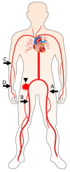
Figure 1
Figure 1
Secondary vascular access sites. Secondary vascular access sites which may be used as ports of entry for a safety wire and delivery of devices for percutaneous repair of femoral artery injury (arrowhead): (A) contralateral femoral artery, (B) ipsilateral distal femoral artery, (C) brachial artery, (D) radial artery.
Figure 2
Figure 2
Secondary VCD failure. An 86-year-old female underwent transfemoral TAVR via the right femoral artery (A). Vessel pre-closure was performed with a Prostar XL device. Control angiogram performed following removal of the 16 Fr sheath and fastening of the Prostar sutures revealed femoral artery perforation (B). A Fluency 9 × 60 mm stent graft which was delivered from the contralateral femoral artery achieved hemostasis (C).
Figure 3
Figure 3
Primary VCD failure. A 90-year-old male with peripheral vascular disease was diagnosed with severe symptomatic aortic stenosis and an abdominal aortic aneurysm. (a) CT angiography revealed an infra-renal aortic aneurysm and diffusely calcified ileofemoral arteries with a minimal lumen diameter of 6.0 mm. (b) A combined TAVR and endovascular aneurysm repair (EVAR) procedure was performed. 5Fr sheaths were inserted into both superficial femoral arteries, through which two 0.014″ safety wires were delivered to the aorta. Pre-closure with Proglide vascular closure devices was performed in the right femoral artery; however, vascular calcification precluded Proglide deployment in the left femoral artery (A). Implantation of a 29 mm Sapien S3 valve (B) was followed by EVAR (C) (safety wires within the femoral arteries marked with arrows). A 9 × 60 mm Fluency stent graft (between arrowheads) was positioned parallel to the 14Fr sheath (arrow) in the left femoral artery prior to sheath removal (D) and deployed following sheath removal (E). Proglide devices achieved vascular closure of the right femoral artery following removal of the 18Fr sheath (F).
Figure 3
Figure 3
Primary VCD failure. A 90-year-old male with peripheral vascular disease was diagnosed with severe symptomatic aortic stenosis and an abdominal aortic aneurysm. (a) CT angiography revealed an infra-renal aortic aneurysm and diffusely calcified ileofemoral arteries with a minimal lumen diameter of 6.0 mm. (b) A combined TAVR and endovascular aneurysm repair (EVAR) procedure was performed. 5Fr sheaths were inserted into both superficial femoral arteries, through which two 0.014″ safety wires were delivered to the aorta. Pre-closure with Proglide vascular closure devices was performed in the right femoral artery; however, vascular calcification precluded Proglide deployment in the left femoral artery (A). Implantation of a 29 mm Sapien S3 valve (B) was followed by EVAR (C) (safety wires within the femoral arteries marked with arrows). A 9 × 60 mm Fluency stent graft (between arrowheads) was positioned parallel to the 14Fr sheath (arrow) in the left femoral artery prior to sheath removal (D) and deployed following sheath removal (E). Proglide devices achieved vascular closure of the right femoral artery following removal of the 18Fr sheath (F).
Figure 4
Figure 4
Use of multiple modalities in a patient with hostile vascular anatomy. An 89-year-old female with severe PVD and severe symptomatic aortic stenosis was referred for TAVR. (a) CT angiography revealed extremely calcified and stenosed ileofemoral arteries, with minimal diameters of 3.5 mm. (b) The TAVR procedure was performed via the left femoral artery, with two safety wires inserted; antegrade via the right brachial artery and retrograde via the left superficial femoral artery (SFA). (A) A 5Fr sheath was inserted into the left SFA (arrow). (B) The left common femoral artery was then punctured and pre-closure with a Proglide device was performed. (C) The left iliac artery was dilated with a 6 mm Shockwave balloon at 6 atmospheres and a 6 mm non-compliant balloon at 24 atmospheres. (D) A 26 mm Evolut Pro valve was delivered over a stiff Lunderquist wire and implanted. (E) An occlusive 7 mm balloon (arrowhead) was delivered via the right brachial artery and inflated within the proximal left iliac artery prior to sheath removal (arrow). (F) Control angiography of the left femoral artery, which was performed via the left SFA following removal of the 16 Fr sheath and fastening of the Proglide sutures, revealed vessel perforation (arrow) and location of both safety wires (arrowheads). (G) Three overlapping stent grafts were implanted within the ileofemoral artery. (H) Repeat angiography confirmed adequate hemostasis. The SFA and brachial sheaths were removed following administration of protamine sulfate.
Figure 5
Figure 5
Flowchart for procedural planning and management of vascular complications. CTA, CT angiography; SW, safety wire; GW, guide wire; PTA, peripheral angioplasty; IVL, intravascular lithotripsy; VCD, vascular closure device; SG, stent graft.

References

    1. Siontis G.C.M., Overtchouk P., Cahill T.J., Modine T., Prendergast B., Praz F., Pilgrim T., Petrinic T., Nikolakopoulou A., Salanti G., et al. Transcatheter aortic valve implantation vs. surgical aortic valve replacement for treatment of symptomatic severe aortic stenosis: An updated meta-analysis. Eur. Heart J. 2019;40:3143–3153. doi: 10.1093/eurheartj/ehz275. - DOI - PubMed
    1. Nguyen V., Willner N., Eltchaninoff H., Burwash I.G., Michel M., Durand E., Gilard M., Dindorf C., Iung B., Cribier A., et al. Trends in aortic valve replacement for aortic stenosis: A French nationwide study. Eur. Heart J. 2022;43:666–679. doi: 10.1093/eurheartj/ehab773. - DOI - PubMed
    1. Morello A., Corcione N., Ferraro P., Cimmino M., Pepe M., Cassese M., Frati G., Biondi-Zoccai G., Giordano A. The best way to transcatheter aortic valve implantation: From standard to new approaches. Int. J. Cardiol. 2021;322:86–94. doi: 10.1016/j.ijcard.2020.08.036. - DOI - PubMed
    1. Eugène M., Duchnowski P., Prendergast B., Wendler O., Laroche C., Monin J.-L., Jobic Y., Popescu B.A., Bax J.J., Vahanian A., et al. Contemporary Management of Severe Symptomatic Aortic Stenosis. J. Am. Coll. Cardiol. 2021;78:2131–2143. doi: 10.1016/j.jacc.2021.09.864. - DOI - PubMed
    1. Costa G., Bieliauskas G., Fukutomi M., Ihlemann N., Søndergaard L., De Backer O. Feasibility and safety of a fully percutaneous transcatheter aortic valve replacement program. Catheter. Cardiovasc. Interv. 2021;97:E418–E424. doi: 10.1002/ccd.29117. - DOI - PubMed

LinkOut - more resources